Intercellular Communication Induces Glycolytic Synchronisation Waves published in PNAS

Intercellular communication induces glycolytic synchronization waves between individually oscillating cells

Intercellular communication induces glycolytic synchronization waves between individually oscillating cells
Martin Mojica-Benavides, David D. van Niekerk, Mite Mijalkov, Jacky L. Snoep, Bernhard Mehlig, Giovanni Volpe, Caroline B. Adiels & Mattias Goksör
PNAS 118(6), e2010075118 (2021)
doi: 10.1073/pnas.2010075118
arXiv: 1909.05187

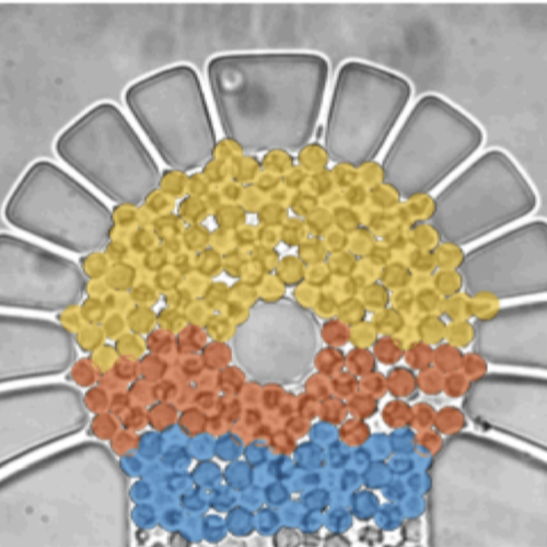
Metabolic oscillations in single cells underlie the mechanisms behind cell synchronization and cell-cell communication. For example, glycolytic oscillations mediated by biochemical communication between cells may synchronize the pulsatile insulin secretion by pancreatic tissue, and a link between glycolytic synchronization anomalies and type-2 diabetes has been hypotesized. Cultures of yeast cells have provided an ideal model system to study synchronization and propagation waves of glycolytic oscillations in large populations. However, the mechanism by which synchronization occurs at individual cell-cell level and overcome local chemical concentrations and heterogenic biological clocks, is still an open question because of experimental limitations in sensitive and specific handling of single cells. Here, we show how the coupling of intercellular diffusion with the phase regulation of individual oscillating cells induce glycolytic synchronization waves. We directly measure the single-cell metabolic responses from yeast cells in a microfluidic environment and characterize a discretized cell-cell communication using graph theory. We corroborate our findings with simulations based on a kinetic detailed model for individual yeast cells. These findings can provide insight into the roles cellular synchronization play in biomedical applications, such as insulin secretion regulation at the cellular level.

Press release on joint research on intercellular communication mechanism by Biological Physics Lab and Soft Matter Lab

The article Intercellular Communication Induces Glycolytic Synchronisation Waves published in PNAS has been featured in the News of the Faculty of Science of Gothenburg University.

Here the links to the press releases:
Swedish: Forskare har knäckt koden för cellkommunikation
English: Researchers have broken the code for cell communication

Delayed correlations improve the reconstruction of the brain connectome published on PLoS ONE

Example of a weighted small-world structural network.

Delayed correlations improve the reconstruction of the brain connectome
Mite Mijalkov, Joana B. Pereira & Giovanni Volpe
PLoS ONE 15(2), e0228334 (2020)
doi: https://doi.org/10.1371/journal.pone.0228334

The brain works as a large-scale complex network, known as the connectome. The strength of the connections between two brain regions in the connectome is commonly estimated by calculating the correlations between their patterns of activation. This approach relies on the assumption that the activation of connected regions occurs together and at the same time. However, there are delays between the activation of connected regions due to excitatory and inhibitory connections. Here, we propose a method to harvest this additional information and reconstruct the structural brain connectome using delayed correlations. This delayed-correlation method correctly identifies 70% to 80% of connections of simulated brain networks, compared to only 5% to 25% of connections detected by the standard methods; this result is robust against changes in the network parameters (small-worldness, excitatory vs. inhibitory connection ratio, weight distribution) and network activation dynamics. The delayed-correlation method predicts more accurately both the global network properties (characteristic path length, global efficiency, clustering coefficient, transitivity) and the nodal network properties (nodal degree, nodal clustering, nodal global efficiency), particularly at lower network densities. We obtain similar results in networks derived from animal and human data. These results suggest that the use of delayed correlations improves the reconstruction of the structural brain connectome and open new possibilities for the analysis of the brain connectome, as well as for other types of networks.

Mite Mijalkov defended his PhD Thesis. Congrats!

Mite Mijalkov defended his PhD Thesis on 24 April 2018 in the Physics Department seminar room (SA240).

Assoc. Prof. Hande Toffoli (Middle-East Technical University), Prof. Tayfun Ozcelik (Bilkent University), Assoc. Prof. Alpan Bek (Middle-East Technical University), Assist. Prof. Seymur Cahangirov (Bilkent Unievrsity) and Assist. Prof. Giovanni Volpe (Bilkent University) will be the thesis committee members.

Thesis title: Graph Theory Study of Complex Networks in the Brain

Thesis abstract: The brain is a large-scale, intricate web of neurons, known as the connectome. By representing the brain as a network i.e. a set of nodes connected by edges, one can study its organization by using concepts from graph theory to evaluate various measures. We have developed BRAPH – BRain Analysis using graPHtheory, a MatLab, object-oriented freeware that facilitates the connectivity analysis of brain networks. BRAPH provides user-friendly interfaces that guide the user through the various steps of the connectivity analysis, such as, calculating adjacency matrices, evaluating global and local measures, performing group comparisons by non-parametric permutations and assessing the communities in a network. Furthermore, using graph theory, we showed that structural MRI undirected networks of stable MCI (sMCI) subjects, late MCI converters (lMCIc), early MCI converters (eMCIc), and AD patients show abnormal organization. This is indicated, at global level, by decreases in clustering and transitivity accompanied by increases in path length and modularity and, at nodal level, by changes in nodal clustering and closeness centrality in patient groups when compared to controls. In samples that do not exhibit differences in the undirected analysis, we propose the usage of directed networks to assess any topological changes due to a neurodegenerative disease. We demonstrate that such changes can be identified in Alzheimer’s and Parkinson’s patients by using directed networks built by delayed correlation coefficients. Finally, we put forward a method that improves the reconstruction of the brain connectome by utilizing the delays in the dynamic behavior of the neurons. We show that this delayed correlationmethod correctly identifies 70% to 80% of the real connections in simulated networks and performs well in the identification of their global and nodal properties.

Name of the PhD programme: Material Science and Nanotechnology Graduate Program
Thesis Advisor  Giovanni Volpe, Department of Physics, Bilkent University

Place: Physics Department seminar room (SA240), Bilkent University
Time: 24 April, 2018, 11:00

BRAPH published in Plos ONE

BRAPH: A graph theory software for the analysis of brain connectivity

BRAPH: A graph theory software for the analysis of brain connectivity
Mite Mijalkov, Ehsan Kakaei, Joana B. Pereira, Eric Westman & Giovanni Volpe
PLoS ONE 12(8), e0178798 (2017)
DOI: 10.1371/journal.pone.0178798
bioRxiv: 106625

The brain is a large-scale complex network whose workings rely on the interaction between its various regions. In the past few years, the organization of the human brain network has been studied extensively using concepts from graph theory, where the brain is represented as a set of nodes connected by edges. This representation of the brain as a connectome can be used to assess important measures that reflect its topological architecture. We have developed a freeware MatLab-based software (BRAPH–BRain Analysis using graPH theory) for connectivity analysis of brain networks derived from structural magnetic resonance imaging (MRI), functional MRI (fMRI), positron emission tomography (PET) and electroencephalogram (EEG) data. BRAPH allows building connectivity matrices, calculating global and local network measures, performing non-parametric permutations for group comparisons, assessing the modules in the network, and comparing the results to random networks. By contrast to other toolboxes, it allows performing longitudinal comparisons of the same patients across different points in time. Furthermore, even though a user-friendly interface is provided, the architecture of the program is modular (object-oriented) so that it can be easily expanded and customized. To demonstrate the abilities of BRAPH, we performed structural and functional graph theory analyses in two separate studies. In the first study, using MRI data, we assessed the differences in global and nodal network topology in healthy controls, patients with amnestic mild cognitive impairment, and patients with Alzheimer’s disease. In the second study, using resting-state fMRI data, we compared healthy controls and Parkin- son’s patients with mild cognitive impairment.

Disrupted Network Topology in Alzheimer published in Cerebral Cortex

Disrupted Network Topology in Patients with Stable and Progressive Mild Cognitive Impairment and Alzheimer’s Disease

Disrupted Network Topology in Patients with Stable and Progressive Mild Cognitive Impairment and Alzheimer’s Disease
Joana B. Pereira, Mite Mijalkov, Ehsan Kakaei, Patricia Mecocci, Bruno Vellas, Magda Tsolaki, Iwona Kłoszewska, Hilka Soininen, Christian Spenger, Simmon Lovestone, Andrew Simmons, Lars-Olof Wahlund, Giovanni Volpe & Eric Westman, AddNeuroMed consortium, for the Alzheimer’s Disease Neuroimaging Initiative
Cerebral Cortex 26(8), 3476—3493 (2016)
DOI: 10.1093/cercor/bhw128

Recent findings suggest that Alzheimer’s disease (AD) is a disconnection syndrome characterized by abnormalities in large- scale networks. However, the alterations that occur in network topology during the prodromal stages of AD, particularly in patients with stable mild cognitive impairment (MCI) and those that show a slow or faster progression to dementia, are still poorly understood. In this study, we used graph theory to assess the organization of structural MRI networks in stable MCI (sMCI) subjects, late MCI converters (lMCIc), early MCI converters (eMCIc), and AD patients from 2 large multicenter cohorts: ADNI and AddNeuroMed. Our findings showed an abnormal global network organization in all patient groups, as reflected by an increased path length, reduced transitivity, and increased modularity compared with controls. In addition, lMCIc, eMCIc, and AD patients showed a decreased path length and mean clustering compared with the sMCI group. At the local level, there were nodal clustering decreases mostly in AD patients, while the nodal closeness centrality detected abnormalities across all patient groups, showing overlapping changes in the hippocampi and amygdala and nonoverlapping changes in parietal, entorhinal, and orbitofrontal regions. These findings suggest that the prodromal and clinical stages of AD are associated with an abnormal network topology.

Engineering of Sensorial Delay published in Phys. Rev. X

Engineering sensorial delay to control phototaxis and emergent collective behaviors

Engineering sensorial delay to control phototaxis and emergent collective behaviors
Mite Mijalkov, Austin McDaniel, Jan Wehr & Giovanni Volpe
Physical Review X 6(1), 011008 (2016)
DOI: 10.1103/PhysRevX.6.011008
arXiv: 1511.04528

Collective motions emerging from the interaction of autonomous mobile individuals play a key role in many phenomena, from the growth of bacterial colonies to the coordination of robotic swarms. For these collective behaviors to take hold, the individuals must be able to emit, sense, and react to signals. When dealing with simple organisms and robots, these signals are necessarily very elementary; e.g., a cell might signal its presence by releasing chemicals and a robot by shining light. An additional challenge arises because the motion of the individuals is often noisy; e.g., the orientation of cells can be altered by Brownian motion and that of robots by an uneven terrain. Therefore, the emphasis is on achieving complex and tunable behaviors from simple autonomous agents communicating with each other in robust ways. Here, we show that the delay between sensing and reacting to a signal can determine the individual and collective long-term behavior of autonomous agents whose motion is intrinsically noisy. We experimentally demonstrate that the collective behavior of a group of phototactic robots capable of emitting a radially decaying light field can be tuned from segregation to aggregation and clustering by controlling the delay with which they change their propulsion speed in response to the light intensity they measure. We track this transition to the underlying dynamics of this system, in particular, to the ratio between the robots’ sensorial delay time and the characteristic time of the robots’ random reorientation. Supported by numerics, we discuss how the same mechanism can be applied to control active agents, e.g., airborne drones, moving in a three-dimensional space. Given the simplicity of this mechanism, the engineering of sensorial delay provides a potentially powerful tool to engineer and dynamically tune the behavior of large ensembles of autonomous mobile agents; furthermore, this mechanism might already be at work within living organisms such as chemotactic cells.

Featured in “Focus: Sensing Delays Control Robot Swarming”, Physics 9, 13 (January 29, 2016)

OTGO published in JOSA B

Computational toolbox for optical tweezers in geometrical optics

Computational toolbox for optical tweezers in geometrical optics
Agnese Callegari, Mite Mijalkov, Burak Gököz & Giovanni Volpe
Journal of the Optical Society of America B 32(5), B11—B19 (2015)
DOI: 10.1364/JOSAB.32.000B11
arXiv: 1402.5439

Optical tweezers have found widespread application in many fields, from physics to biology. Here, we explain in detail how optical forces and torques can be described within the geometrical optics approximation, and we show that this approximation provides reliable results in agreement with experiments for particles whose characteristic dimensions are larger than the wavelength of the trapping light. Furthermore, we provide an object-oriented software package implemented in MATLAB for the calculation of optical forces and torques in the geometrical optics regime: Optical Tweezers in Geometrical Optics (OTGO). We provide all source codes for OTGO as well as documentation and code examples—e.g., standard optical tweezers, optical tweezers with elon- gated particles, the windmill effect, and Kramers transitions between two optical traps—necessary to enable users to effectively employ it in their research.

Sorting of Chiral Microswimmers published in Soft Matter

Sorting of chiral microswimmers

Sorting of chiral microswimmers (Cover article)
Mite Mijalkov & Giovanni Volpe
Soft Matter 9(28), 6376—6381 (2013)
DOI: 10.1039/C3SM27923E
arXiv: 1212.6504

Microscopic swimmers, e.g., chemotactic bacteria and cells, are capable of directed motion by exerting a force on their environment. For asymmetric microswimmers, e.g., bacteria, spermatozoa and many artificial active colloidal particles, a torque is also present leading to circular motion (in two dimensions) and to helicoidal motion (in three dimensions) with a well-defined chirality. Here, we demonstrate with numerical simulations in two dimensions how the chirality of circular motion couples to chiral features present in the microswimmer environment. Levogyre and dextrogyre microswimmers as small as 50 nm can be separated and selectively trapped in chiral flowers of ellipses. Patterned microchannels can be used as funnels to rectify the microswimmer motion, as sorters to separate microswimmers based on their linear and angular velocities, and as sieves to trap microswimmers with specific parameters. We also demonstrate that these results can be extended to helicoidal motion in three dimensions.