BRAPH 2 Genesis enables swift creation of custom, reproducible software distributions—tackling the growing complexity of neuroscience by streamlining analysis across diverse data types and workflows. (Image by B. Zufiria-Gerbolés and Y.-W. Chang.)BRAPH 2: a flexible, open-source, reproducible, community-oriented, easy-to-use framework for network analyses in neurosciences
Yu-Wei Chang, Blanca Zufiria-Gerbolés, Pablo Emiliano Gómez-Ruiz, Anna Canal-Garcia, Hang Zhao, Mite Mijalkov, Joana Braga Pereira, Giovanni Volpe
bioRxiv: 10.1101/2025.04.11.648455
As network analyses in neuroscience continue to grow in both complexity and size, flexible methods are urgently needed to provide unbiased, reproducible insights into brain function. BRAPH 2 is a versatile, open-source framework that meets this challenge by offering streamlined workflows for advanced statistical models and deep learning in a community-oriented environment. Through its Genesis compiler, users can build specialized distributions with custom pipelines, ensuring flexibility and scalability across diverse research domains. These powerful capabilities will ensure reproducibility and accelerate discoveries in neuroscience.
Memory capacity in aging. A Brain reservoir computing architecture with uniform random signals applied to all nodes. (Image from the article.)Computational memory capacity predicts aging and cognitive decline
Mite Mijalkov, Ludvig Storm, Blanca Zufiria-Gerbolés, Dániel Veréb, Zhilei Xu, Anna Canal-Garcia, Jiawei Sun, Yu-Wei Chang, Hang Zhao, Emiliano Gómez-Ruiz, Massimiliano Passaretti, Sara Garcia-Ptacek, Miia Kivipelto, Per Svenningsson, Henrik Zetterberg, Heidi Jacobs, Kathy Lüdge, Daniel Brunner, Bernhard Mehlig, Giovanni Volpe, Joana B. Pereira
Nature Communications 16, 2748 (2025)
doi: 10.1038/s41467-025-57995-0
Memory is a crucial cognitive function that deteriorates with age. However, this ability is normally assessed using cognitive tests instead of the architecture of brain networks. Here, we use reservoir computing, a recurrent neural network computing paradigm, to assess the linear memory capacities of neural-network reservoirs extracted from brain anatomical connectivity data in a lifespan cohort of 636 individuals. The computational memory capacity emerges as a robust marker of aging, being associated with resting-state functional activity, white matter integrity, locus coeruleus signal intensity, and cognitive performance. We replicate our findings in an independent cohort of 154 young and 72 old individuals. By linking the computational memory capacity of the brain network with cognition, brain function and integrity, our findings open new pathways to employ reservoir computing to investigate aging and age-related disorders.
The book Deep Learning Crash Course, authored by Giovanni Volpe, Benjamin Midtvedt, Jesús Pineda, Henrik Klein Moberg, Harshith Bachimanchi, Joana B. Pereira, and Carlo Manzo, has been published online by No Starch Press in July 2024.
Citation
Giovanni Volpe, Benjamin Midtvedt, Jesús Pineda, Henrik Klein Moberg, Harshith Bachimanchi, Joana B. Pereira, and Carlo Manzo. Deep Learning Crash Course. No Starch Press.
ISBN-13: 9781718503922
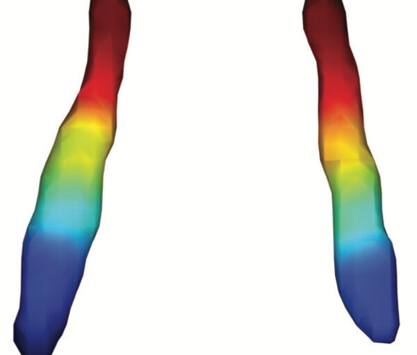
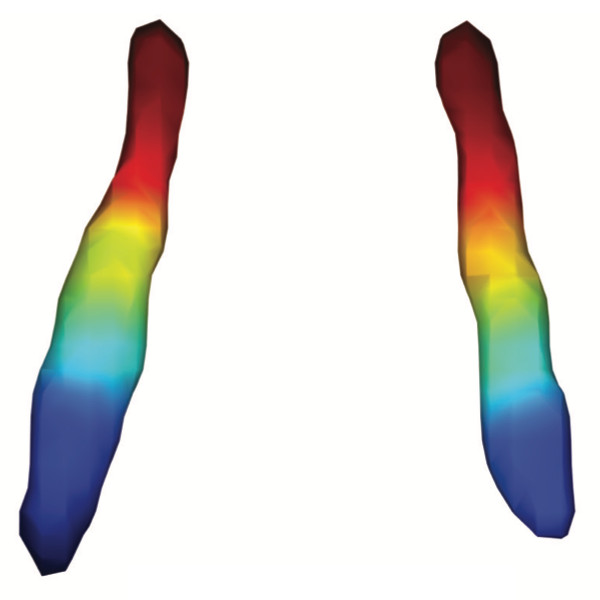
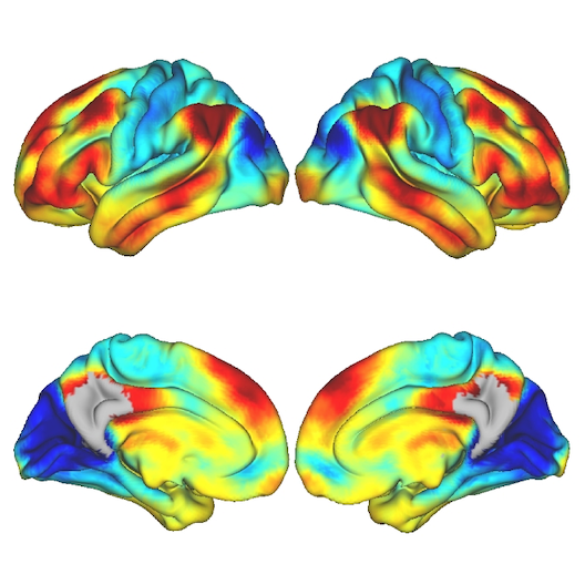
Average functional gradients of the locus coeruleus in the CamCAN 3T dataset. (Image from the publication.)Age-related differences in the functional topography of the locus coeruleus and their implications for cognitive and affective functions
Dániel Veréb, Mite Mijalkov, Anna Canal-Garcia, Yu-Wei Chang, Emiliano Gomez-Ruiz, Blanca Zufiria Gerboles, Miia Kivipelto, Per Svenningsson, Henrik Zetterberg, Giovanni Volpe, Matthew Betts, Heidi IL Jacobs, Joana B Pereira
eLife 12, RP87188 (2023)
doi: 10.7554/eLife.87188.3
The locus coeruleus (LC) is an important noradrenergic nucleus that has recently attracted a lot of attention because of its emerging role in cognitive and psychiatric disorders. Although previous histological studies have shown that the LC has heterogeneous connections and cellular features, no studies have yet assessed its functional topography in vivo, how this heterogeneity changes over aging, and whether it is associated with cognition and mood. Here, we employ a gradient-based approach to characterize the functional heterogeneity in the organization of the LC over aging using 3T resting-state fMRI in a population-based cohort aged from 18 to 88 years of age (Cambridge Centre for Ageing and Neuroscience cohort, n=618). We show that the LC exhibits a rostro-caudal functional gradient along its longitudinal axis, which was replicated in an independent dataset (Human Connectome Project [HCP] 7T dataset, n=184). Although the main rostro-caudal direction of this gradient was consistent across age groups, its spatial features varied with increasing age, emotional memory, and emotion regulation. More specifically, a loss of rostral-like connectivity, more clustered functional topography, and greater asymmetry between right and left LC gradients was associated with higher age and worse behavioral performance. Furthermore, participants with higher-than-normal Hospital Anxiety and Depression Scale (HADS) ratings exhibited alterations in the gradient as well, which manifested in greater asymmetry. These results provide an in vivo account of how the functional topography of the LC changes over aging, and imply that spatial features of this organization are relevant markers of LC-related behavioral measures and psychopathology.
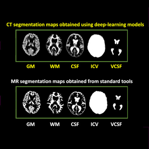
Imaging-based volumetric measures. (Image by the Authors of the manuscript.)CT-based volumetric measures obtained through deep learning: Association with biomarkers of neurodegeneration
Meera Srikrishna, Nicholas J. Ashton, Alexis Moscoso, Joana B. Pereira, Rolf A. Heckemann, Danielle van Westen, Giovanni Volpe, Joel Simrén, Anna Zettergren, Silke Kern, Lars-Olof Wahlund, Bibek Gyanwali, Saima Hilal, Joyce Chong Ruifen, Henrik Zetterberg, Kaj Blennow, Eric Westman, Christopher Chen, Ingmar Skoog, Michael Schöll
Alzheimer’s & Dementia 20, 629–640 (2024)
arXiv: 2401.06260
doi: 10.1002/alz.13445
INTRODUCTION
Cranial computed tomography (CT) is an affordable and widely available imaging modality that is used to assess structural abnormalities, but not to quantify neurodegeneration. Previously we developed a deep-learning–based model that produced accurate and robust cranial CT tissue classification.
MATERIALS AND METHODS
We analyzed 917 CT and 744 magnetic resonance (MR) scans from the Gothenburg H70 Birth Cohort, and 204 CT and 241 MR scans from participants of the Memory Clinic Cohort, Singapore. We tested associations between six CT-based volumetric measures (CTVMs) and existing clinical diagnoses, fluid and imaging biomarkers, and measures of cognition.
RESULTS
CTVMs differentiated cognitively healthy individuals from dementia and prodromal dementia patients with high accuracy levels comparable to MR-based measures. CTVMs were significantly associated with measures of cognition and biochemical markers of neurodegeneration.
DISCUSSION
These findings suggest the potential future use of CT-based volumetric measures as an informative first-line examination tool for neurodegenerative disease diagnostics after further validation.
Illustration of resting state network activity. (Image by the Authors of the manuscript.)Peripheral inflammatory subgroup differences in anterior Default Mode network and multiplex functional network topology are associated with cognition in psychosis
Paulo Lizano, Chelsea Kiely, Mite Mijalkov, Shashwath A. Meda, Sarah K. Keedy, Dung Hoang, Victor Zeng, Olivia Lutz, Joana B. Pereira, Elena I. Ivleva, Giovanni Volpe, Yanxun Xu, Adam M. Lee, Leah H. Rubin, S Kristian Hill, Brett A. Clementz, Carol A. Tamminga, Godfrey D. Pearlson, John A. Sweeney, Elliot S. Gershon, Matcheri S. Keshavan, Jeffrey R. Bishop
Brain Behavior and Immunity, 114, 3-15 (2023)
doi: 10.1016/j.bbi.2023.07.014
Introduction
High-inflammation subgroups of patients with psychosis demonstrate cognitive deficits and neuroanatomical alterations. Systemic inflammation assessed using IL-6 and C-reactive protein may alter functional connectivity within and between resting-state networks, but the cognitive and clinical implications of these alterations remain unknown. We aim to determine the relationships of elevated peripheral inflammation subgroups with resting-state functional networks and cognition in psychosis spectrum disorders.
Methods
Serum and resting-state fMRI were collected from psychosis probands (schizophrenia, schizoaffective, psychotic bipolar disorder) and healthy controls (HC) from the B-SNIP1 (Chicago site) study who were stratified into inflammatory subgroups based on factor and cluster analyses of 13 cytokines (HC Low n = 32, Proband Low n = 65, Proband High n = 29). Nine resting-state networks derived from independent component analysis were used to assess functional and multilayer connectivity. Inter-network connectivity was measured using Fisher z-transformation of correlation coefficients. Network organization was assessed by investigating networks of positive and negative connections separately, as well as investigating multilayer networks using both positive and negative connections. Cognition was assessed using the Brief Assessment of Cognition in Schizophrenia. Linear regressions, Spearman correlations, permutations tests and multiple comparison corrections were used for analyses in R.
Results
Anterior default mode network (DMNa) connectivity was significantly reduced in the Proband High compared to Proband Low (Cohen’s d = -0.74, p = 0.002) and HC Low (d = -0.85, p = 0.0008) groups. Inter-network connectivity between the DMNa and the right-frontoparietal networks was lower in Proband High compared to Proband Low (d = -0.66, p = 0.004) group. Compared to Proband Low, the Proband High group had lower negative (d = 0.54, p = 0.021) and positive network (d = 0.49, p = 0.042) clustering coefficient, and lower multiplex network participation coefficient (d = -0.57, p = 0.014). Network findings in high inflammation subgroups correlate with worse verbal fluency, verbal memory, symbol coding, and overall cognition.
Conclusion
These results expand on our understanding of the potential effects of peripheral inflammatory signatures and/or subgroups on network dysfunction in psychosis and how they relate to worse cognitive performance. Additionally, the novel multiplex approach taken in this study demonstrated how inflammation may disrupt the brain’s ability to maintain healthy co-activation patterns between the resting-state networks while inhibiting certain connections between them.
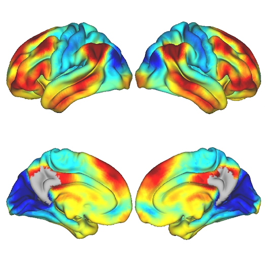
Spatial maps depicting the strongest connections from the medial parietal cortex to other cortical and subcortical areas in the PREVENT-AD cohort. (Reproduced from the publication.)Functional gradients of the medial parietal cortex in a healthy cohort with family history of sporadic Alzheimer’s disease
Dániel Veréb, Mite Mijalkov, Yu-Wei Chang, Anna Canal-Garcia, Emiliano Gomez-Ruis, Anne Maass, Sylvia Villeneuve, Giovanni Volpe Joana B. Pereira
Alzheimer’s Research & Therapy 15, 82 (2023)
doi: 10.1186/s13195-023-01228-3
Background
The medial parietal cortex is an early site of pathological protein deposition in Alzheimer’s disease (AD). Previous studies have identified different subregions within this area; however, these subregions are often heterogeneous and disregard individual differences or subtle pathological alterations in the underlying functional architecture. To address this limitation, here we measured the continuous connectivity gradients of the medial parietal cortex and assessed their relationship with cerebrospinal fluid (CSF) biomarkers, ApoE ε4 carriership and memory in asymptomatic individuals at risk to develop AD.
Methods
Two hundred sixty-three cognitively normal participants with a family history of sporadic AD who underwent resting-state and task-based functional MRI using encoding and retrieval tasks were included from the PREVENT-AD cohort. A novel method for characterizing spatially continuous patterns of functional connectivity was applied to estimate functional gradients in the medial parietal cortex during the resting-state and task-based conditions. This resulted in a set of nine parameters that described the appearance of the gradient across different spatial directions. We performed correlation analyses to assess whether these parameters were associated with CSF biomarkers of phosphorylated tau181 (p-tau), total tau (t-tau), and amyloid-ß1-42 (Aß). Then, we compared the spatial parameters between ApoE ε4 carriers and noncarriers, and evaluated the relationship between these parameters and memory.
Results
Alterations involving the superior part of the medial parietal cortex, which was connected to regions of the default mode network, were associated with higher p-tau, t-tau levels as well as lower Aß/p-tau levels during the resting-state condition (p < 0.01). Similar alterations were found in ApoE ε4 carriers compared to non-carriers (p < 0.003). In contrast, lower immediate memory scores were associated with changes in the middle part of the medial parietal cortex, which was connected to inferior temporal and posterior parietal regions, during the encoding task (p = 0.001). No results were found when using conventional connectivity measures.
Conclusions
Functional alterations in the medial parietal gradients are associated with CSF AD biomarkers, ApoE e4 carriership, and lower memory in an asymptomatic cohort with a family history of sporadic AD, suggesting that functional gradients are sensitive to subtle changes associated with early AD stages.
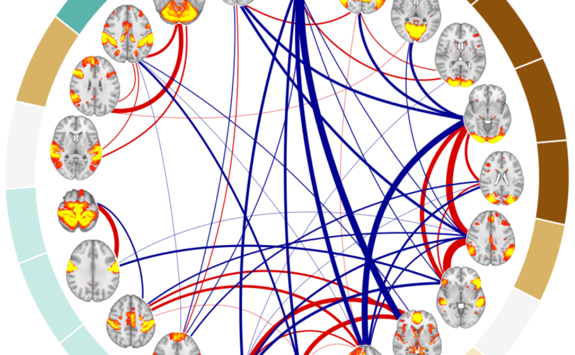
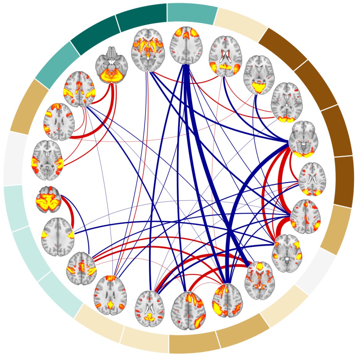
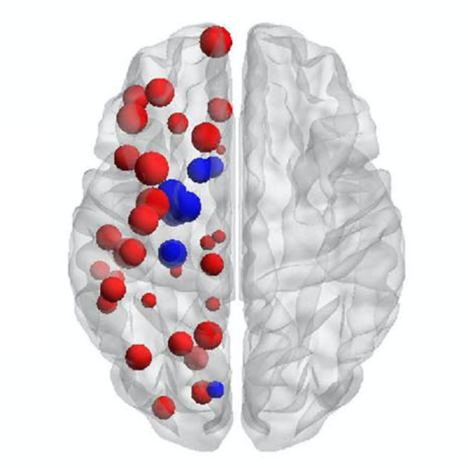
Example of the 21 resting-state networks used as nodes and their positive (red) and negative connections (blue) for one of 140 the subjects included in the analyses. (Image by the Authors of the manuscript.)Sex differences in multilayer functional network topology over the course of aging in 37543 UK Biobank participants
Mite Mijalkov, Dániel Veréb, Oveis Jamialahmadi, Anna Canal-Garcia, Emiliano Gómez-Ruiz, Didac Vidal-Piñeiro, Stefano Romeo, Giovanni Volpe, Joana B. Pereira
Network Neuroscience 1-40 (2022)
doi: 10.1162/netn_a_00286
medRxiv: 10.1101/2022.03.08.22272089
Aging is a major risk factor for cardiovascular and neurodegenerative disorders, with considerable societal and economic implications. Healthy aging is accompanied by changes in functional connectivity between and within resting-state functional networks, which have been associated with cognitive decline. However, there is no consensus on the impact of sex on these age-related functional trajectories. Here, we show that multilayer measures provide crucial information on the interaction between sex and age on network topology, allowing for better assessment of cognitive, structural, and cardiovascular risk factors that have been shown to differ between men and women, as well as providing additional insights into the genetic influences on changes in functional connectivity that occur during aging. In a large cross-sectional sample of 37543 individuals from the UK Biobank cohort, we demonstrate that such multilayer measures that capture the relationship between positive and negative connections are more sensitive to sex-related changes in the whole-brain connectivity patterns and their topological architecture throughout aging, when compared to standard connectivity and topological measures. Our findings indicate that multilayer measures contain previously unknown information on the relationship between sex and age, which opens up new avenues for research into functional brain connectivity in aging.
Comparison of cluster-specific covariance matrixes with node strength. (Image by the Authors.)Multi-cohort and longitudinal Bayesian clustering study of stage and subtype in Alzheimer’s disease
Konstantinos Poulakis, Joana B. Pereira, J.-Sebastian Muehlboeck, Lars-Olof Wahlund, Örjan Smedby, Giovanni Volpe, Colin L. Masters, David Ames, Yoshiki Niimi, Takeshi Iwatsubo, Daniel Ferreira, Eric Westman, Japanese Alzheimer’s Disease Neuroimaging Initiative & Australian Imaging, Biomarkers and Lifestyle study
Nature Communications 13, 4566 (2022)
doi: 10.1038/s41467-022-32202-6
Understanding Alzheimer’s disease (AD) heterogeneity is important for understanding the underlying pathophysiological mechanisms of AD. However, AD atrophy subtypes may reflect different disease stages or biologically distinct subtypes. Here we use longitudinal magnetic resonance imaging data (891 participants with AD dementia, 305 healthy control participants) from four international cohorts, and longitudinal clustering to estimate differential atrophy trajectories from the age of clinical disease onset. Our findings (in amyloid-β positive AD patients) show five distinct longitudinal patterns of atrophy with different demographical and cognitive characteristics. Some previously reported atrophy subtypes may reflect disease stages rather than distinct subtypes. The heterogeneity in atrophy rates and cognitive decline within the five longitudinal atrophy patterns, potentially expresses a complex combination of protective/risk factors and concomitant non-AD pathologies. By alternating between the cross-sectional and longitudinal understanding of AD subtypes these analyses may allow better understanding of disease heterogeneity.
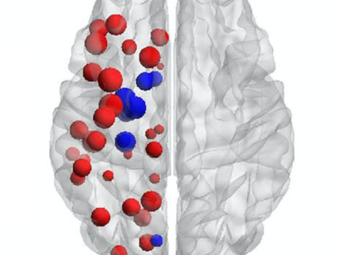
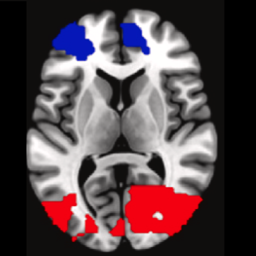
Particular of the brain in the group comparison analysis. (Image by the Authors.)Unraveling Parkinson’s disease heterogeneity using subtypes based on multimodal data
Franziska Albrecht, Konstantinos Poulakis, Malin Freidle, Hanna Johansson, Urban Ekman, Giovanni Volpe, Eric Westman, Joana B. Pereira, Erika Franzén
Parkinsonism and Related Disorders 102, 19-29 (2022)
doi: 10.1016/j.parkreldis.2022.07.014
Background
Parkinson’s disease (PD) is a clinically and neuroanatomically heterogeneous neurodegenerative disease characterized by different subtypes. To this date, no studies have used multimodal data that combines clinical, motor, cognitive and neuroimaging assessments to identify these subtypes, which may provide complementary, clinically relevant information. To address this limitation, we subtyped participants with mild-moderate PD based on a rich, multimodal dataset of clinical, cognitive, motor, and neuroimaging variables.
Methods
Cross-sectional data from 95 PD participants from our randomized EXPANd (EXercise in PArkinson’s disease and Neuroplasticity) controlled trial were included. Participants were subtyped using clinical, motor, and cognitive assessments as well as structural and resting-state MRI data. Subtyping was done by random forest clustering. We extracted information about the subtypes by inspecting their neuroimaging profiles and descriptive statistics.
Results
Our multimodal subtyping analysis yielded three PD subtypes: a motor-cognitive subtype characterized by widespread alterations in brain structure and function as well as impairment in motor and cognitive abilities; a cognitive dominant subtype mainly impaired in cognitive function that showed frontoparietal structural and functional changes; and a motor dominant subtype impaired in motor variables without any brain alterations. Motor variables were most important for the subtyping, followed by gray matter volume in the right medial postcentral gyrus.
Conclusions
Three distinct PD subtypes were identified in our multimodal dataset. The most important features to subtype PD participants were motor variables in addition to structural MRI in the sensorimotor region. These findings have the potential to improve our understanding of PD heterogeneity, which in turn can lead to personalized interventions and rehabilitation.