Harshith Bachimanchi won best early career researcher presentation award at ETAI 2023, San Diego

The three award winners. From left to right: Mite Mijalkov, Harshith Bachimanchi, Marie Drouhin. (Photo by G. Volpe.)
Harshith Bachimanchi won the best early career researcher presentation (gold) award at Emerging Topics in Artificial Intelligence (ETAI) 2023 held in San Diego, California, USA, from 20 – 24 August 2023. The award, consisting of an invitation to a part of a perspective article of AI in neurosciences, is offered by the organisers of the conference, and SPIE Optics + Photonics.

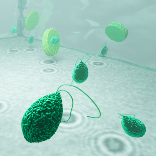
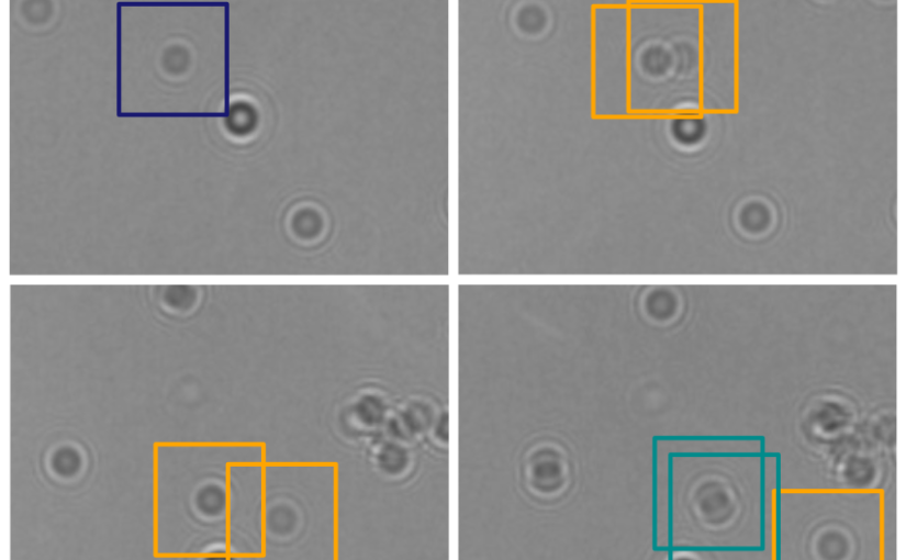
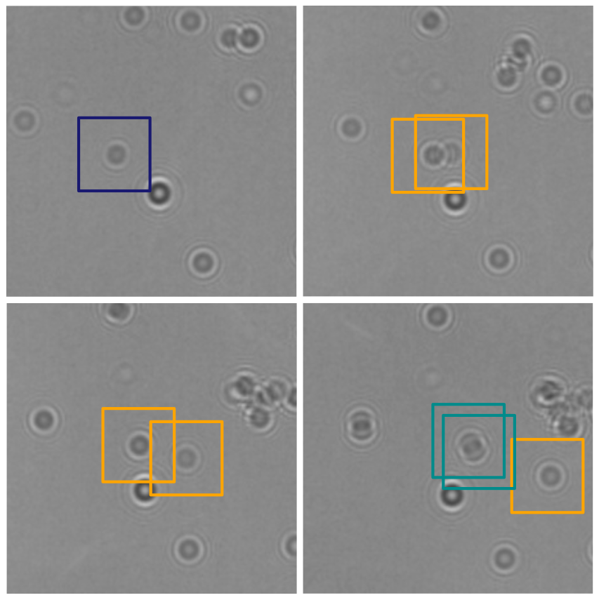
In this work, Harshith presented his recent work on combining holographic microscopy and deep learning to study the marine microplankton. He demonstrated that the combination of holographic microscopy and deep learning can be used to follow the marine microorganisms throughout their lifespan, continuously measuring their three-dimensional positions and dry mass. The deep-learning algorithms circumvent the computationally intensive processing of holographic data and allow rapid measurements over extended periods of time. This enables to reliably estimate growth rates, both in terms of dry mass increase and cell divisions, as well as to measure trophic interactions between species such as predation events. Studying individual interactions in idealized small systems provides insights that help us understand microbial food webs and ultimately larger-scale processes. He exemplified this by showing detailed descriptions of micro-zooplankton feeding events, cell divisions, and long-term monitoring of single cells from division to division.

The article related to this presentation can be found at the following link: Microplankton life histories revealed by holographic microscopy and deep learning.

Award certificate of Harshith Bachimanchi. (Provided by H. Bachimanchi.)
Harshith Bachimanchi receives the award from Joana B. Pereira. (Photo by G. Volpe.)
The three award winners. From left to right: Mite Mijalkov, Harshith Bachimanchi, Marie Drouhin. (Photo by G. Volpe.)

Presentation by H. Bachimanchi at SPIE-ETAI, San Diego, 23 August 2023

Planktons imaged under a holographic microscope. (Illustration by J. Heuschele.)
Decoding microplankton life through holographic microscopy and deep learning
Harshith Bachimanchi
23 August 2023, 8:45 AM PDT

The marine microbial food web plays a central role in the global carbon cycle. However, our mechanistic understanding of the ocean is biased toward its larger constituents, while rates and biomass fluxes in the microbial food web are mainly inferred from indirect measurements and ensemble averages. Yet, resolution at the level of the individual microplankton is required to advance our understanding of the microbial food web. Here, we demonstrate that, by combining holographic microscopy with deep learning, we can follow microplanktons throughout their lifespan, continuously measuring their three-dimensional position and dry mass. The deep-learning algorithms circumvent the computationally intensive processing of holographic data and allow rapid measurements over extended time periods. This permits us to reliably estimate growth rates, both in terms of dry mass increase and cell divisions, as well as to measure trophic interactions between species such as predation events. The individual resolution provides information about selectivity, individual feeding rates, and handling times for individual microplanktons. The method is particularly useful to detail the rates and routes of organic matter transfer in micro-zooplankton, the most important and least known group of primary consumers in the oceans. Studying individual interactions in idealized small systems provides insights that help us understand microbial food webs and ultimately larger-scale processes. We exemplify this by detailed descriptions of micro-zooplankton feeding events, cell divisions, and long-term monitoring of single cells from division to division.

The article related to this presentation can be found at the following link: Microplankton life histories revealed by holographic microscopy and deep learning.

Presentation by H. Bachimanchi at XVII International Congress of the Spanish Biophysical Society, Castelldefels, 30 June 2023

Planktons imaged under a holographic microscope. (Illustration by J. Heuschele.)
Bringing microplankton into focus: Deep learning meets holographic microscopy
Harshith Bachimanchi
30 June 2023, 12:40 CEST

The marine microbial food web plays a central role in the global carbon cycle. However, our mechanistic understanding of the ocean is biased toward its larger constituents, while rates and biomass fluxes in the microbial food web are mainly inferred from indirect measurements and ensemble averages. Yet, resolution at the level of the individual microplankton is required to advance our understanding of the microbial food web. Here, we demonstrate that, by combining holographic microscopy with deep learning, we can follow microplanktons throughout their lifespan, continuously measuring their three-dimensional position and dry mass. The deep-learning algorithms circumvent the computationally intensive processing of holographic data and allow rapid measurements over extended time periods. This permits us to reliably estimate growth rates, both in terms of dry mass increase and cell divisions, as well as to measure trophic interactions between species such as predation events. The individual resolution provides information about selectivity, individual feeding rates, and handling times for individual microplanktons. The method is particularly useful to detail the rates and routes of organic matter transfer in micro-zooplankton, the most important and least known group of primary consumers in the oceans. Studying individual interactions in idealized small systems provides insights that help us understand microbial food webs and ultimately larger-scale processes. We exemplify this by detailed descriptions of micro-zooplankton feeding events, cell divisions, and long-term monitoring of single cells from division to division.

The article related to this presentation can be found at the following link: Microplankton life histories revealed by holographic microscopy and deep learning.

Presentation by H. Bachimanchi at AI for Scientific Data Analysis, Gothenburg, 1 June 2023

Planktons imaged under a holographic microscope. (Illustration by J. Heuschele.)
Quantitative microplankton tracking by holographic microscopy and deep learning
Harshith Bachimanchi
1 June 2023, 15:00 CEST

The marine microbial food web plays a central role in the global carbon cycle. However, our mechanistic understanding of the ocean is biased toward its larger constituents, while rates and biomass fluxes in the microbial food web are mainly inferred from indirect measurements and ensemble averages. Yet, resolution at the level of the individual microplankton is required to advance our understanding of the microbial food web. Here, we demonstrate that, by combining holographic microscopy with deep learning, we can follow microplanktons throughout their lifespan, continuously measuring their three-dimensional position and dry mass. The deep-learning algorithms circumvent the computationally intensive processing of holographic data and allow rapid measurements over extended time periods. This permits us to reliably estimate growth rates, both in terms of dry mass increase and cell divisions, as well as to measure trophic interactions between species such as predation events. The individual resolution provides information about selectivity, individual feeding rates, and handling times for individual microplanktons. The method is particularly useful to detail the rates and routes of organic matter transfer in micro-zooplankton, the most important and least known group of primary consumers in the oceans. Studying individual interactions in idealized small systems provides insights that help us understand microbial food webs and ultimately larger-scale processes. We exemplify this by detailed descriptions of micro-zooplankton feeding events, cell divisions, and long-term monitoring of single cells from division to division.

The article related to this presentation can be found at the following link: Microplankton life histories revealed by holographic microscopy and deep learning.

Presentation by H. Bachimanchi at Signals in the Sea mini-symposium, Lund University, 12 May 2023

Planktons imaged under a holographic microscope. (Illustration by J. Heuschele.)
Deep learning in plankton ecology
Harshith Bachimanchi
Presentation at Biology department, Lund University
12 May 2023, 13:00 CEST

In this mini-symposium organised by Dr. Erik Selander at Lund University, I have spoken about our recent work with marine microplankton, where we have combined holographic microscopy with deep learning to measure the ‘dry’ mass and 3D swimming dynamics of different species of planktons. The article related to this presentation can be found at the following here: Microplankton life histories revealed by holographic microscopy and deep learning.

The presentation was followed by discussions with Prof. Karin Rengefors group at Lund university on the topic of application of AI based methods for various kinds of studies in phytoplankton ecology and evolution.

Geometric deep learning reveals the spatiotemporal fingerprint of microscopic motion published in Nature Machine Intelligence

Input graph structure including a redundant number of edges. (Image by J. Pineda.)
Geometric deep learning reveals the spatiotemporal fingerprint of microscopic motion
Jesús Pineda, Benjamin Midtvedt, Harshith Bachimanchi, Sergio Noé, Daniel Midtvedt, Giovanni Volpe, Carlo Manzo
Nature Machine Intelligence 5, 71–82 (2023)
arXiv: 2202.06355
doi: 10.1038/s42256-022-00595-0

The characterization of dynamical processes in living systems provides important clues for their mechanistic interpretation and link to biological functions. Thanks to recent advances in microscopy techniques, it is now possible to routinely record the motion of cells, organelles, and individual molecules at multiple spatiotemporal scales in physiological conditions. However, the automated analysis of dynamics occurring in crowded and complex environments still lags behind the acquisition of microscopic image sequences. Here, we present a framework based on geometric deep learning that achieves the accurate estimation of dynamical properties in various biologically-relevant scenarios. This deep-learning approach relies on a graph neural network enhanced by attention-based components. By processing object features with geometric priors, the network is capable of performing multiple tasks, from linking coordinates into trajectories to inferring local and global dynamic properties. We demonstrate the flexibility and reliability of this approach by applying it to real and simulated data corresponding to a broad range of biological experiments.

Single-shot self-supervised object detection in microscopy published in Nature Communications

LodeSTAR tracks the plankton Noctiluca scintillans. (Image by the Authors of the manuscript.)
Single-shot self-supervised particle tracking
Benjamin Midtvedt, Jesús Pineda, Fredrik Skärberg, Erik Olsén, Harshith Bachimanchi, Emelie Wesén, Elin K. Esbjörner, Erik Selander, Fredrik Höök, Daniel Midtvedt, Giovanni Volpe
Nature Communications 13, 7492 (2022)
arXiv: 2202.13546
doi: 10.1038/s41467-022-35004-y

Object detection is a fundamental task in digital microscopy, where machine learning has made great strides in overcoming the limitations of classical approaches. The training of state-of-the-art machine-learning methods almost universally relies on vast amounts of labeled experimental data or the ability to numerically simulate realistic datasets. However, experimental data are often challenging to label and cannot be easily reproduced numerically. Here, we propose a deep-learning method, named LodeSTAR (Localization and detection from Symmetries, Translations And Rotations), that learns to detect microscopic objects with sub-pixel accuracy from a single unlabeled experimental image by exploiting the inherent roto-translational symmetries of this task. We demonstrate that LodeSTAR outperforms traditional methods in terms of accuracy, also when analyzing challenging experimental data containing densely packed cells or noisy backgrounds. Furthermore, by exploiting additional symmetries we show that LodeSTAR can measure other properties, e.g., vertical position and polarizability in holographic microscopy.

Recent eLife article on plankton tracking gets featured on Swedish national radio

Planktons imaged under a holographic microscope. (Illustration by J. Heuschele.)
The article Microplankton life histories revealed by holographic microscopy and deep learning gets featured on Vetenskapradion Nyheter (Science radio) operated by Sveriges Radio (Swedish national radio) on November 7, 2022.

The short audio feature (Hologram hjälper forskare att förstå plankton) which highlights the important results of the paper (in Swedish) is now available for public listening.

Vetenskapradion Nyheter airs daily news, reports and in-depth discussions about latest research.

Press release on Microplankton life histories revealed by holographic microscopy and deep learning

Planktons imaged under a holographic microscope. (Illustration by J. Heuschele.)
The article Microplankton life histories revealed by holographic microscopy and deep learning has been featured in the news of University of Gothenburg (in English & Swedish) and in the press release of eLife (in English).

The study, now published in eLife, and co-written by researchers at the Soft Matter Lab of the Department of Physics at the University of Gothenburg, demonstrates how the combination of holographic microscopy and deep learning provides a strong complimentary tool in marine microbial ecology. The research allows quantitative assessments of microplankton feeding behaviours, and biomass increase throughout the cell cycle from generation to generation.

The study is featured also in eLife digest.

Here are the links to the press releases:
Researchers combine microscopy with AI to characterise marine microbial food web (eLife, English)
Holographic microscopy provides insights into the life of microplankton (GU, English)
Hologram ger insyn i planktonens liv (GU, Swedish)
The secret lives of microbes (eLife digest)

Microplankton life histories revealed by holographic microscopy and deep learning published in eLife

Tracking of microplankton by holographic optical microscopy and deep learning. (Image by H. Bachimanchi.)
Microplankton life histories revealed by holographic microscopy and deep learning
Harshith Bachimanchi, Benjamin Midtvedt, Daniel Midtvedt, Erik Selander, and Giovanni Volpe
eLife 11, e79760 (2022)
arXiv: 2202.09046
doi: 10.7554/eLife.79760

The marine microbial food web plays a central role in the global carbon cycle. Our mechanistic understanding of the ocean, however, is biased towards its larger constituents, while rates and biomass fluxes in the microbial food web are mainly inferred from indirect measurements and ensemble averages. Yet, resolution at the level of the individual microplankton is required to advance our understanding of the oceanic food web. Here, we demonstrate that, by combining holographic microscopy with deep learning, we can follow microplanktons throughout their lifespan, continuously measuring their three dimensional position and dry mass. The deep learning algorithms circumvent the computationally intensive processing of holographic data and allow rapid measurements over extended time periods. This permits us to reliably estimate growth rates, both in terms of dry mass increase and cell divisions, as well as to measure trophic interactions between species such as predation events. The individual resolution provides information about selectivity, individual feeding rates and handling times for individual microplanktons. This method is particularly useful to explore the flux of carbon through micro-zooplankton, the most important and least known group of primary consumers in the global oceans. We exemplify this by detailed descriptions of micro-zooplankton feeding events, cell divisions, and long term monitoring of single cells from division to division.