Max Haraldsson joined the Soft Matter Lab on 9 February 2026.
Max is a master student in Biomedical Engineering at Chalmers University of Technology.
Together with his classmate Anton Widengård, he will be conducting his Master’s thesis at the Soft Matter Lab in collaboration with IFLAI, with Jesús Pineda as supervisor.
Max’s and Anton’s project is about evaluating and efficiently adapting pre-trained deep learning models for cell segmentation and tracking.
Cover of the PhD thesis. (Image by F. Skärberg)Fredrik Skärberg defended his PhD thesis on January 29th, 2026. Congrats!
The defense took place in FB, Institutionen för fysik, Origovägen 6b, Göteborg, at 09:00.
Title: From Light to Data Using Deep Learning for Quantitative Microscopy
Abstract: Quantitative microscopy aims to measure physical properties of microscopic particles from optical images, but weak and complex signals often make this difficult. This thesis explores how computational methods, especially deep learning guided by physical understanding, can improve particle detection and characterization in microscopy.
The work introduces new approaches for locating and tracking particles, extends these ideas to three-dimensional and label-free imaging, and reviews practical analysis workflows. It further shows how combining complementary imaging techniques can enhance nanoparticle measurements and how deep learning can recover three-dimensional structural information from microscopy images.
Overall, this thesis strengthens the connection between optical measurements and quantitative particle information, expanding the potential of label-free microscopy for biological and nanoscale studies.
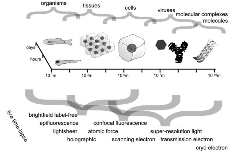
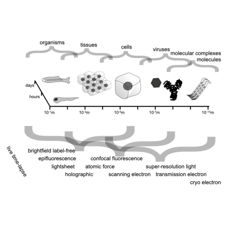
Spatio-temporal spectrum diagram of microscopy techniques and their applications. (Image by the Authors of the manuscript.)Roadmap on Deep Learning for Microscopy
Giovanni Volpe, Carolina Wählby, Lei Tian, Michael Hecht, Artur Yakimovich, Kristina Monakhova, Laura Waller, Ivo F. Sbalzarini, Christopher A. Metzler, Mingyang Xie, Kevin Zhang, Isaac C.D. Lenton, Halina Rubinsztein-Dunlop, Daniel Brunner, Bijie Bai, Aydogan Ozcan, Daniel Midtvedt, Hao Wang, Nataša Sladoje, Joakim Lindblad, Jason T. Smith, Marien Ochoa, Margarida Barroso, Xavier Intes, Tong Qiu, Li-Yu Yu, Sixian You, Yongtao Liu, Maxim A. Ziatdinov, Sergei V. Kalinin, Arlo Sheridan, Uri Manor, Elias Nehme, Ofri Goldenberg, Yoav Shechtman, Henrik K. Moberg, Christoph Langhammer, Barbora Špačková, Saga Helgadottir, Benjamin Midtvedt, Aykut Argun, Tobias Thalheim, Frank Cichos, Stefano Bo, Lars Hubatsch, Jesus Pineda, Carlo Manzo, Harshith Bachimanchi, Erik Selander, Antoni Homs-Corbera, Martin Fränzl, Kevin de Haan, Yair Rivenson, Zofia Korczak, Caroline Beck Adiels, Mite Mijalkov, Dániel Veréb, Yu-Wei Chang, Joana B. Pereira, Damian Matuszewski, Gustaf Kylberg, Ida-Maria Sintorn, Juan C. Caicedo, Beth A Cimini, Muyinatu A. Lediju Bell, Bruno M. Saraiva, Guillaume Jacquemet, Ricardo Henriques, Wei Ouyang, Trang Le, Estibaliz Gómez-de-Mariscal, Daniel Sage, Arrate Muñoz-Barrutia, Ebba Josefson Lindqvist, Johanna Bergman
Journal of Physics: Photonics 8, 012501 (2026)
arXiv: 2303.03793
doi: 10.1088/2515-7647/ae0fd1
Through digital imaging, microscopy has evolved from primarily being a means for visual observation of life at the micro- and nano-scale, to a quantitative tool with ever-increasing resolution and throughput. Artificial intelligence, deep neural networks, and machine learning (ML) are all niche terms describing computational methods that have gained a pivotal role in microscopy-based research over the past decade. This Roadmap encompasses key aspects of how ML is applied to microscopy image data, with the aim of gaining scientific knowledge by improved image quality, automated detection, segmentation, classification and tracking of objects, and efficient merging of information from multiple imaging modalities. We aim to give the reader an overview of the key developments and an understanding of possibilities and limitations of ML for microscopy. It will be of interest to a wide cross-disciplinary audience in the physical sciences and life sciences.
Cover of the PhD thesis. (Image by B. García)Berenice García defended her PhD thesis on January 28th, 2026. Congrats!
The defense took place in PJ, Institutionen för fysik, Origovägen 6b, Göteborg, at 09:00.
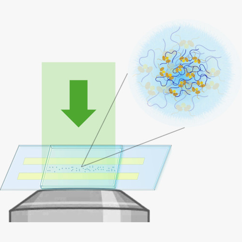
Title: Quantitative Optical Microscopy of Microscale Soft Matter Systems
Abstract: Many biological and soft-matter particles operate at sizes below the diffraction limit and scatter light only weakly, making them hard to study with conventional microscopy. This thesis introduces two complementary, label-free interferometric methods that enable single-particle characterization across the meso–microscale. By combining optical scattering, off-axis holography, and particle tracking, these approaches quantify size, refractive index, internal structure, and mobility of individual rigid nanoparticles and soft biomolecular condensates. Together, this work provides new tools for probing the physical principles of nanoscale soft matter and phase-separated biological assemblies.
Supervisor: Daniel Midtvedt Examiner: Bernhard Mehlig Opponent: Balpreet Singh Ahluwalia Committee: Per Augustsson, Arrate Muñoz Barrutia, Alexandra Stubelius Alternate board member: Kristian Gustafsson
Cover of the PhD thesis. (Image by Hula King, https://www.behance.net/hulaking)Yu-Wei Chang defended his PhD thesis on January 23rd, 2026. Congrats!
The defense will take place in SB-H7 lecture hall, SB-Building, Institutionen för fysik, Johanneberg Campus, Göteborg, at 13:00.
Title: A Unified Software-Generating Framework for Biological Data Analysis
Abstract: Biological data analysis relies heavily on software, but as projects grow it becomes hard to keep code, interfaces, and tests aligned, and to reuse methods without rewriting them. This thesis presents Genesis, which generates runnable modules, GUIs, and unit tests from a single human-readable .gen.m description of each analysis component. By maintaining a central library of these descriptions, analyses can be recombined for new questions while staying consistent. Four studies across neuroimaging, light-sheet microscopy, and plant Raman spectroscopy show the framework is reusable and extensible across domains.
Hang Zhao, supervised by Giovanni Volpe and Joana Pereira, will present his halftime seminar under the topic “Brain connectome revealed neuro-degenerative disease” on 9-10 am, 22nd Jan. 2026 in Nexus and through Zoom (https://gu-se.zoom.us/j/7726618257). The seminar starts from his presentation about the past and planned project, followed by a discussion and questions by his opponent, Professor Mattias Göksor.
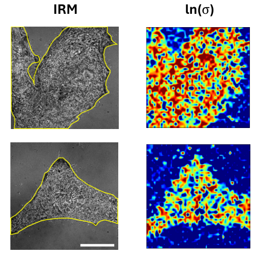
Quantifying the spatiotemporal forces, affinities, and dissipative costs of cellular-scale non-equilibrium processes from experimental data and localizing it in space and time remain a significant open challenge. Here, I explore how principles from stochastic thermodynamics, combined with machine learning techniques, offer a promising approach to addressing this issue. I will present preliminary results from experiments on fluctuating cell membranes and simulations of non-equilibrium systems in stationary and time-dependently driven states. These studies reveal potential strategies for localizing entropy production in experimental biophysical contexts while also highlighting key challenges and limitations that must be addressed.
Eduard Andrei Duta Costache started his PhD at the Physics Department of the University of Gothenburg on the 19th of January 2026.
Eduard has a double Master’s degree in Artificial Intelligence from the University of Alicante (Spain) and in Machine Learning & Data Mining from Jean Monnet University (France).
During the course of his PhD, as part of the GREENS MSCA Doctoral Network, he will focus on developing AI frameworks to model and optimize the lifecycle of micro-robotic platforms.
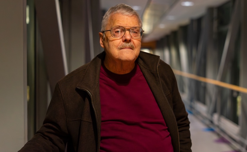
Steven B. Smith. (Photo by A. Ciarlo)Steven Smith will visit the Soft Matter Lab from 17 to 28 January 2026.
Steven brings years of expertise from the laboratory of Professor Carlos Bustamante, where they pioneered the use and development of optical tweezers. As the main developer of the widely used ‘minitweezers’ instrument, which today is used by dozens of research groups, he has helped shape the field of single-molecule biophysics on a global scale. We look forward to his visit, during which he will work on refining cutting-edge single-molecule measurement techniques.