News

The Cognitive Connectome in Healthy Aging published in Frontiers in Aging Neuroscience

Age-independent cognitive connectome in the whole cohort.
The Cognitive Connectome in Healthy Aging
Eloy Garcia-Cabello, Lissett Gonzalez-Burgos, Joana B. Pereira, Juan Andres Hernández-Cabrera, Eric Westman, Giovanni Volpe, José Barroso, & Daniel Ferreira
Front. Aging Neurosci. 13, 530 (2021)
doi: 10.3389/fnagi.2021.694254

Objectives: Cognitive aging has been extensively investigated using both univariate and multivariate analyses. Sophisticated multivariate approaches such as graph theory could potentially capture unknown complex associations between multiple cognitive variables. The aim of this study was to assess whether cognition is organized into a structure that could be called the “cognitive connectome,” and whether such connectome differs between age groups.

Methods: A total of 334 cognitively unimpaired individuals were stratified into early-middle-age (37–50 years, n = 110), late-middle-age (51–64 years, n = 106), and elderly (65–78 years, n = 118) groups. We built cognitive networks from 47 cognitive variables for each age group using graph theory and compared the groups using different global and nodal graph measures.

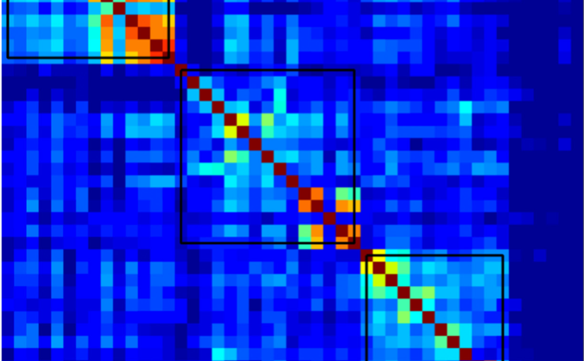
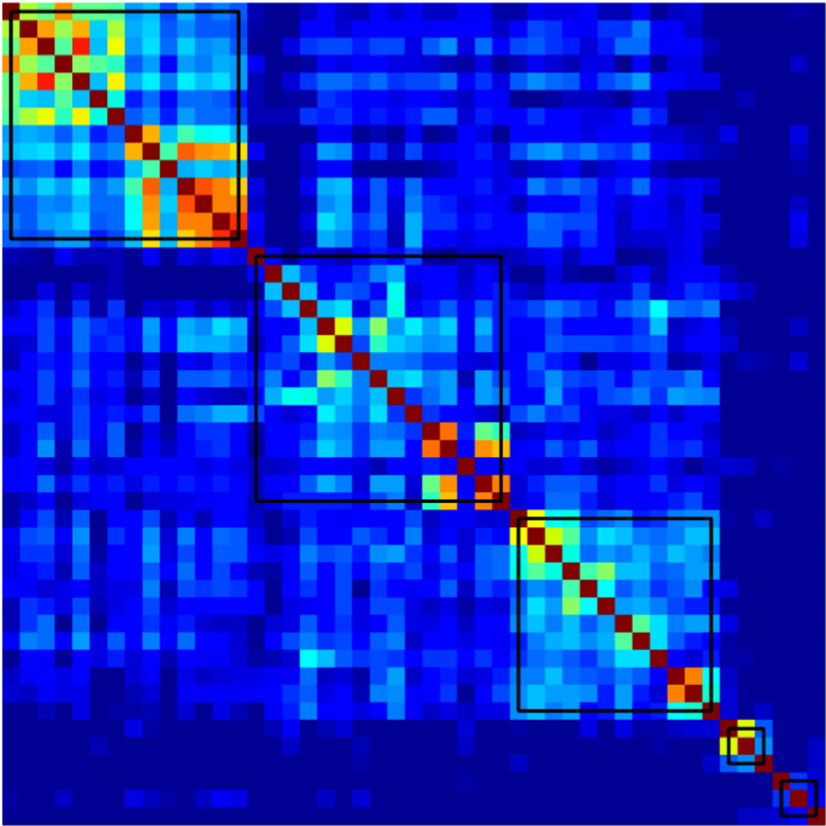
Results: We identified a cognitive connectome characterized by five modules: verbal memory, visual memory—visuospatial abilities, procedural memory, executive—premotor functions, and processing speed. The elderly group showed reduced transitivity and average strength as well as increased global efficiency compared with the early-middle-age group. The late-middle-age group showed reduced global and local efficiency and modularity compared with the early-middle-age group. Nodal analyses showed the important role of executive functions and processing speed in explaining the differences between age groups.

Conclusions: We identified a cognitive connectome that is rather stable during aging in cognitively healthy individuals, with the observed differences highlighting the important role of executive functions and processing speed. We translated the connectome concept from the neuroimaging field to cognitive data, demonstrating its potential to advance our understanding of the complexity of cognitive aging.

Enhanced prediction of atrial fibrillation and mortality among patients with congenital heart disease using nationwide register-based medical hospital data and neural networks published in European Heart Journal – Digital Health

Neural network prediction of mortality and atrial fibrillation. (Image taken from the article’s graphical abstract.)
Enhanced prediction of atrial fibrillation and mortality among patients with congenital heart disease using nationwide register based medical hospital data and neural networks
Kok Wai Giang, Saga Helgadottir, Mikael Dellborg, Giovanni Volpe, Zacharias Mandalenakis
European Heart Journal – Digital Health (2021)
doi: 10.1093/ehjdh/ztab065

Aims: To improve short-and long-term predictions of mortality and atrial fibrillation (AF) among patients with congenital heart disease (CHD) from a nationwide population using neural networks (NN).

Methods and results: The Swedish National Patient Register and the Cause of Death Register were used to identify all patients with CHD born from 1970 to 2017. A total of 71 941 CHD patients were identified and followed-up from birth until the event or end of study in 2017. Based on data from a nationwide population, a NN model was obtained to predict mortality and AF. Logistic regression (LR) based on the same data was used as a baseline comparison. Of 71 941 CHD patients, a total of 5768 died (8.02%) and 995 (1.38%) developed AF over time with a mean follow-up time of 16.47 years (standard deviation 12.73 years). The performance of NN models in predicting the mortality and AF was higher than the performance of LR regardless of the complexity of the disease, with an average area under the receiver operating characteristic of >0.80 and >0.70, respectively. The largest differences were observed in mortality and complexity of CHD over time.

Conclusion: We found that NN can be used to predict mortality and AF on a nationwide scale using data that are easily obtainable by clinicians. In addition, NN showed a high performance overall and, in most cases, with better performance for prediction as compared with more traditional regression methods.

Directed Brain Connectivity Identifies Widespread Functional Network Abnormalities in Parkinson’s Disease published in Cerebral Cortex

Differences between controls and PD participants in nodal network measures. (Image taken from the article.)
Directed Brain Connectivity Identifies Widespread Functional Network Abnormalities in Parkinson’s Disease
Mite Mijalkov, Giovanni Volpe, Joana B Pereira
Cerebral Cortex, bhab237 (2021)
doi: 10.1093/cercor/bhab237

Parkinson’s disease (PD) is a neurodegenerative disorder characterized by topological abnormalities in large-scale functional brain networks, which are commonly analyzed using undirected correlations in the activation signals between brain regions. This approach assumes simultaneous activation of brain regions, despite previous evidence showing that brain activation entails causality, with signals being typically generated in one region and then propagated to other ones. To address this limitation, here, we developed a new method to assess whole-brain directed functional connectivity in participants with PD and healthy controls using antisymmetric delayed correlations, which capture better this underlying causality. Our results show that whole-brain directed connectivity, computed on functional magnetic resonance imaging data, identifies widespread differences in the functional networks of PD participants compared with controls, in contrast to undirected methods. These differences are characterized by increased global efficiency, clustering, and transitivity combined with lower modularity. Moreover, directed connectivity patterns in the precuneus, thalamus, and cerebellum were associated with motor, executive, and memory deficits in PD participants. Altogether, these findings suggest that directional brain connectivity is more sensitive to functional network differences occurring in PD compared with standard methods, opening new opportunities for brain connectivity analysis and development of new markers to track PD progression.

Extracting quantitative biological information from brightfield cell images using deep learning featured in AIP SciLight

The article Extracting quantitative biological information from brightfield cell images using deep learning
has been featured in: “Staining Cells Virtually Offers Alterative Approach to Chemical Dyes”, AIP SciLight (July 23, 2021).

Scilight showcases the most interesting research across the physical sciences published in AIP Publishing Journals.

Scilight is published weekly (52 issues per year) by AIP Publishing.

Microscopic Metavehicles Powered and Steered by Embedded Optical Metasurfaces published in Nature Nanotechnology

Metavehicles.
Microscopic Metavehicles Powered and Steered by Embedded Optical Metasurfaces
Daniel Andrén, Denis G. Baranov, Steven Jones, Giovanni Volpe, Ruggero Verre, Mikael Käll
Nat. Nanotechnol. (2021)
doi: 10.1038/s41565-021-00941-0
arXiv: 2012.10205

Nanostructured dielectric metasurfaces offer unprecedented opportunities to manipulate light by imprinting an arbitrary phase gradient on an impinging wavefront. This has resulted in the realization of a range of flat analogues to classical optical components, such as lenses, waveplates and axicons. However, the change in linear and angular optical momentum associated with phase manipulation also results in previously unexploited forces and torques that act on the metasurface itself. Here we show that these optomechanical effects can be utilized to construct optical metavehicles – microscopic particles that can travel long distances under low-intensity plane-wave illumination while being steered by the polarization of the incident light. We demonstrate movement in complex patterns, self-correcting motion and an application as transport vehicles for microscopic cargoes, which include unicellular organisms. The abundance of possible optical metasurfaces attests to the prospect of developing a wide variety of metavehicles with specialized functional behaviours.

Extracting quantitative biological information from bright-field cell images using deep learning published in Biophysics Reviews

Virtually-stained generated image for lipid-droplet.
Extracting quantitative biological information from bright-field cell images using deep learning
Saga Helgadottir, Benjamin Midtvedt, Jesús Pineda, Alan Sabirsh, Caroline B. Adiels, Stefano Romeo, Daniel Midtvedt, Giovanni Volpe
Biophysics Rev. 2, 031401 (2021)
arXiv: 2012.12986
doi: 10.1063/5.0044782

Quantitative analysis of cell structures is essential for biomedical and pharmaceutical research. The standard imaging approach relies on fluorescence microscopy, where cell structures of interest are labeled by chemical staining techniques. However, these techniques are often invasive and sometimes even toxic to the cells, in addition to being time-consuming, labor-intensive, and expensive. Here, we introduce an alternative deep-learning-powered approach based on the analysis of bright-field images by a conditional generative adversarial neural network (cGAN). We show that this approach can extract information from the bright-field images to generate virtually-stained images, which can be used in subsequent downstream quantitative analyses of cell structures. Specifically, we train a cGAN to virtually stain lipid droplets, cytoplasm, and nuclei using bright-field images of human stem-cell-derived fat cells (adipocytes), which are of particular interest for nanomedicine and vaccine development. Subsequently, we use these virtually-stained images to extract quantitative measures about these cell structures. Generating virtually-stained fluorescence images is less invasive, less expensive, and more reproducible than standard chemical staining; furthermore, it frees up the fluorescence microscopy channels for other analytical probes, thus increasing the amount of information that can be extracted from each cell.

Presentation by L. Pérez at ELS 2021, 13 July 2021

Laura Pérez presented the work “FORMA and BEFORE: expanding applications of optical tweezers” at the ELS conference (online) on the 13th of July.

The main objective of the Electromagnetic and Light Scattering Conference (ELS) is to bring together scientists and engineers studying various aspects of light scattering and to provide a relaxed academic atmosphere for in-depth discussions of theoretical advances, measurements, and applications.

FORMA allows to identify and characterize all the equilibrium points in a force field generated by a speckle pattern.
FORMA and BEFORE: Expanding Applications of Optical Tweezers. Laura Pérez Garcia, Martin Selin, Alejandro V. Arzola, Giovanni Volpe, Alessandro Magazzù, Isaac Pérez Castillo.
ELS 2021
Date: 13 July 2021
Time: 15:45 (CEST)

Abstract: 
FORMA (force reconstruction via maximum-likelihood-estimator analysis) addresses the need to measure the force fields acting on microscopic particles. Compared to alternative established methods, FORMA is faster, simpler, more accurate, and more precise. Furthermore, FORMA can also measure non-conservative and out-of-equilibrium force fields. Here, after a brief introduction to FORMA, I will present its use, advantages, and limitations. I will conclude with the most recent work where we exploit Bayesian inference to expand FORMA’s scope of application.

Classification, inference and segmentation of anomalous diffusion with recurrent neural networks published in Journal of Physics A: Mathematical and Theoretical

RANDI architecture to classify the model underlying anomalous diffusion.
Classification, inference and segmentation of anomalous diffusion with recurrent neural networks
Aykut Argun, Giovanni Volpe, Stefano Bo
J. Phys. A: Math. Theor. 54 294003 (2021)
doi: 10.1088/1751-8121/ac070a
arXiv: 2104.00553

Countless systems in biology, physics, and finance undergo diffusive dynamics. Many of these systems, including biomolecules inside cells, active matter systems and foraging animals, exhibit anomalous dynamics where the growth of the mean squared displacement with time follows a power law with an exponent that deviates from 1. When studying time series recording the evolution of these systems, it is crucial to precisely measure the anomalous exponent and confidently identify the mechanisms responsible for anomalous diffusion. These tasks can be overwhelmingly difficult when only few short trajectories are available, a situation that is common in the study of non-equilibrium and living systems. Here, we present a data-driven method to analyze single anomalous diffusion trajectories employing recurrent neural networks, which we name RANDI. We show that our method can successfully infer the anomalous exponent, identify the type of anomalous diffusion process, and segment the trajectories of systems switching between different behaviors. We benchmark our performance against the state-of-the art techniques for the study of single short trajectories that participated in the Anomalous Diffusion (AnDi) challenge. Our method proved to be the most versatile method, being the only one to consistently rank in the top 3 for all tasks proposed in the AnDi challenge.

Colloquium by G. Volpe at TU-Darmstadt, 18 June 2021, Online

Deep learning for particle tracking. (Image by Aykut Argun)
Deep learning for microscopy, optical trapping, and active matter

Giovanni Volpe
Colloquium
(online at) TU-Darmstadt, Germany
18 June 2021, 14:00 CEST

After a brief overview of artificial intelligence, machine learning and deep learning, I will present a series of recent works in which we have employed deep learning for applications in photonics and active matter.
In particular, I will explain how we employed deep learning to enhance digital video microscopy, to estimate the properties of anomalous diffusion, to characterize microscopic force fields, to improve the calculation of optical forces, and to characterize nanoparticles.
Finally, I will provide an outlook for the application of deep learning in photonics and active matter.

Saga Helgadottir defended her PhD Thesis in Physics on June 16, 2021. Congrats!

Digital video microscopy enhanced by deep learning
Saga Helgadottir defended her PhD Thesis in Physics on June 16, 2021. Congrats!

The disputation took place at 9 a.m. digitally via Zoom. A link to the Zoom meeting was published the day before dissertation on the GU website.

Title:  Deep Learning Applications – From image analysis to medical diagnosis

Abstract:
Deep learning is a subcategory of machine learning and artificial intelligence. Instead of using explicit rules to perform a desired task as in standard algorithmic approaches, machine-learning algorithms autonomously learn from data to determine the rules for the task at hand. The idea of deep learning has been around since the 1950s but was for a long time limited by available computational power and amount of training data. Once overcome these problems, in recent years, deep learning has made great advances in solving various problems.

In this thesis, I show how deep learning can be applied in image analysis and medical diagnosis, while outperforming standard algorithmic methods and simpler machine-learning methods. I begin with showing that a convolutional neural network trained with simulated particle images is able to track experimental single particles, even in poor illumination conditions. I then show how this inspired the development of an all-in-one software to design, train and validate deep-learning solutions for digital microscopy, from particle tracking and characterization in 2D and 3D to the segmentation, characterization and counting of biological cells and image transformation. I show that this software package can be further used to develop a generative adversarial neural network to virtually stain brightfield images of cells, replacing the traditional chemical staining for a downstream analysis of biological features. I then move on from applications in microscopy and image analysis to show the potential of deep learning in medical diagnosis. I show that dense neural networks perform better than simpler machine-learning algorithm and the clinical standard in the diagnosis of a genetic disease and in the prediction of short- and long-term morbidity in patients with congenital-heart-disease. At last, I have shown that a neural network- powered strategy for testing and isolating individuals adapts to the parameters of a disease outbreak achieves an epidemic containment.

The interdisciplinary nature of the work in this thesis has allowed the application of new technologies developed in the field of physics to solve problems in the fields of biology and biomedicine, as well as overcoming barriers for the continued revolutionization of deep learning in microscopy.

Thesishttp://hdl.handle.net/2077/67506

Supervisor: Giovanni Volpe
Examiner: Bernhard Mehlig
Opponent: Carolina Wählby
Committee: Marj Tonini, Maria Garcia-Parajo, Alexander Rohrbach

Screenshots from Saga Helgadottir’s PhD Thesis defense.

PhD Opponent’s presentation.
PhD Thesis presentation: Saga Helgadottir, Giovanni Volpe (Supervisor), Raimund Feifel (GU Physics), Carolina Wählby (Opponent), Marj Tonini (Committee member), Maria Garcia-Parajo (Committee member), Måns Henningson (GU Physics Department Chair), Alexander Rohrbach (Committee member).
PhD Thesis presentation.
PhD Thesis presentation front slide.
PhD Thesis presentation content slide (1).
PhD Thesis presentation content slide (2).
PhD Thesis presentation conclusion slide.
Screenshot from the discussion (1).
Screenshot from the discussion (2).
Screenshot from the discussion (3).