Extracting quantitative biological information from bright-field cell images using deep learning published in Biophysics Reviews

Virtually-stained generated image for lipid-droplet.
Extracting quantitative biological information from bright-field cell images using deep learning
Saga Helgadottir, Benjamin Midtvedt, Jesús Pineda, Alan Sabirsh, Caroline B. Adiels, Stefano Romeo, Daniel Midtvedt, Giovanni Volpe
Biophysics Rev. 2, 031401 (2021)
arXiv: 2012.12986
doi: 10.1063/5.0044782

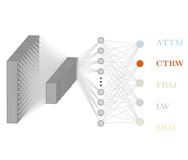
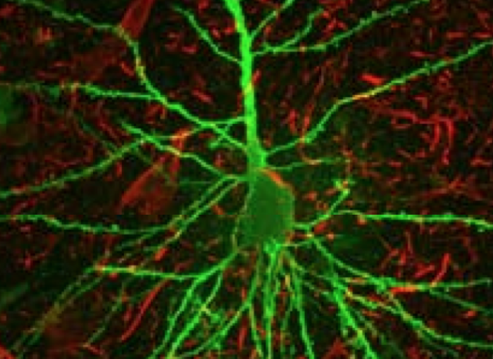
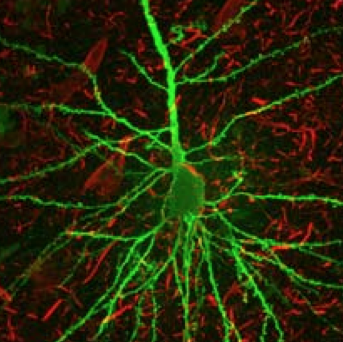
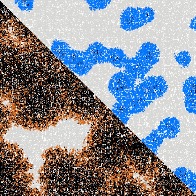
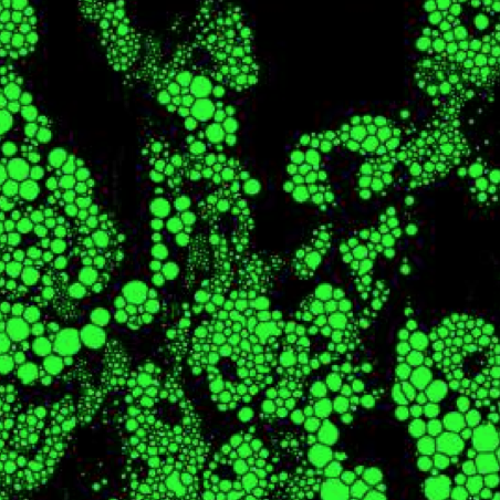
Quantitative analysis of cell structures is essential for biomedical and pharmaceutical research. The standard imaging approach relies on fluorescence microscopy, where cell structures of interest are labeled by chemical staining techniques. However, these techniques are often invasive and sometimes even toxic to the cells, in addition to being time-consuming, labor-intensive, and expensive. Here, we introduce an alternative deep-learning-powered approach based on the analysis of bright-field images by a conditional generative adversarial neural network (cGAN). We show that this approach can extract information from the bright-field images to generate virtually-stained images, which can be used in subsequent downstream quantitative analyses of cell structures. Specifically, we train a cGAN to virtually stain lipid droplets, cytoplasm, and nuclei using bright-field images of human stem-cell-derived fat cells (adipocytes), which are of particular interest for nanomedicine and vaccine development. Subsequently, we use these virtually-stained images to extract quantitative measures about these cell structures. Generating virtually-stained fluorescence images is less invasive, less expensive, and more reproducible than standard chemical staining; furthermore, it frees up the fluorescence microscopy channels for other analytical probes, thus increasing the amount of information that can be extracted from each cell.

Classification, inference and segmentation of anomalous diffusion with recurrent neural networks published in Journal of Physics A: Mathematical and Theoretical

RANDI architecture to classify the model underlying anomalous diffusion.
Classification, inference and segmentation of anomalous diffusion with recurrent neural networks
Aykut Argun, Giovanni Volpe, Stefano Bo
J. Phys. A: Math. Theor. 54 294003 (2021)
doi: 10.1088/1751-8121/ac070a
arXiv: 2104.00553

Countless systems in biology, physics, and finance undergo diffusive dynamics. Many of these systems, including biomolecules inside cells, active matter systems and foraging animals, exhibit anomalous dynamics where the growth of the mean squared displacement with time follows a power law with an exponent that deviates from 1. When studying time series recording the evolution of these systems, it is crucial to precisely measure the anomalous exponent and confidently identify the mechanisms responsible for anomalous diffusion. These tasks can be overwhelmingly difficult when only few short trajectories are available, a situation that is common in the study of non-equilibrium and living systems. Here, we present a data-driven method to analyze single anomalous diffusion trajectories employing recurrent neural networks, which we name RANDI. We show that our method can successfully infer the anomalous exponent, identify the type of anomalous diffusion process, and segment the trajectories of systems switching between different behaviors. We benchmark our performance against the state-of-the art techniques for the study of single short trajectories that participated in the Anomalous Diffusion (AnDi) challenge. Our method proved to be the most versatile method, being the only one to consistently rank in the top 3 for all tasks proposed in the AnDi challenge.

Colloquium by G. Volpe at TU-Darmstadt, 18 June 2021, Online

Deep learning for particle tracking. (Image by Aykut Argun)
Deep learning for microscopy, optical trapping, and active matter

Giovanni Volpe
Colloquium
(online at) TU-Darmstadt, Germany
18 June 2021, 14:00 CEST

After a brief overview of artificial intelligence, machine learning and deep learning, I will present a series of recent works in which we have employed deep learning for applications in photonics and active matter.
In particular, I will explain how we employed deep learning to enhance digital video microscopy, to estimate the properties of anomalous diffusion, to characterize microscopic force fields, to improve the calculation of optical forces, and to characterize nanoparticles.
Finally, I will provide an outlook for the application of deep learning in photonics and active matter.

Dendritic spines are lost in clusters in patients with Alzheimer’s disease published in Scientific Report

Combined confocal microscopy picture showing a neuron with a soma free of PHF-tau.
Dendritic spines are lost in clusters in patients with Alzheimer’s disease
Mite Mijalkov, Giovanni Volpe, Isabel Fernaud-Espinosa, Javier DeFelipe, Joana B. Pereira, Paula Merino-Serrais
Sci. Rep. 11, 12350 (2021)
doi: 10.1038/s41598-021-91726-x
biorXiv: 10.1101/2020.10.20.346718

Alzheimer’s disease (AD) is a progressive neurodegenerative disorder characterized by a deterioration of neuronal connectivity. The pathological accumulation of tau protein in neurons is one of the hallmarks of AD and has been connected to the loss of dendritic spines of pyramidal cells, which are the major targets of cortical excitatory synapses and key elements in memory storage. However, the detailed mechanisms underlying the loss of dendritic spines in patients with AD are still unclear. Here, comparing dendrites with and without tau pathology of AD patients, we show that the presence of tau pathology determines the loss of dendritic spines in blocks, ruling out alternative models where spine loss occurs randomly. Since memory storage has been associated with synaptic clusters, the present results provide a new insight into the mechanisms by which tau drives synaptic damage in AD, paving the way to memory deficits by altering spine organization.

Presentation by L. Natali at Spatial Data Science 2020, 11 June 2021

Comparison of different evolution regimes of disease spreading: free evolution (bottom left half) vs network strategy (top right half). (Image by Laura Natali.)
Improving epidemic testing and containment strategies using machine learning. 
Laura Natali, Saga Helgadottir, Onofrio M. Maragò, Giovanni Volpe.
Submitted to SDS2020
Date: 11 June
Time: 16:15 (CEST)

Abstract: 
Containment of epidemic outbreaks entails great societal and economic costs.  Cost-effective containment strategies rely on efficiently identifying infected individuals, making the best possible use of the available testing resources. Therefore, quickly identifying the optimal testing strategy is of critical importance. Here, we demonstrate that machine learning can be used to identify which individuals are most beneficial to test, automatically and dynamically adapting the testing strategy to the characteristics of the disease outbreak. Specifically, we simulate an outbreak using the archetypal susceptible-infectious-recovered (SIR) model and we use data about the first confirmed cases to train a neural network that learns to make predictions about the rest of the population. Using these prediction, we manage to contain the outbreak more effectively and more quickly than with standard approaches. Furthermore, we demonstrate how this method can be used also when there is a possibility of reinfection (SIRS model) to efficiently eradicate an endemic disease.

Seminar by G. Volpe at Vi2, University of Uppsala, 17 May 2021

Deep learning for particle tracking. (Image by Aykut Argun.)
Quantitative Digital Microscopy with Deep Learning
Giovanni Volpe
Seminar
Vi2 Seminar (Visual Information and Interaction)
University of Uppsala
17 May 2021, 14:15 CEST
Online

Video microscopy has a long history of providing insights and breakthroughs for a broad range of disciplines, from physics to biology. Image analysis to extract quantitative information from video microscopy data has traditionally relied on algorithmic approaches, which are often difficult to implement, time consuming, and computationally expensive. Recently, alternative data-driven approaches using deep learning have greatly improved quantitative digital microscopy, potentially offering automatized, accurate, and fast image analysis. However, the combination of deep learning and video microscopy remains underutilized primarily due to the steep learning curve involved in developing custom deep-learning solutions. To overcome this issue, we introduce a software, DeepTrack 2.0, to design, train and validate deep-learning solutions for digital microscopy. We use it to exemplify how deep learning can be employed for a broad range of applications, from particle localization, tracking and characterization to cell counting and classification. Thanks to its user-friendly graphical interface, DeepTrack 2.0 can be easily customized for user-specific applications, and, thanks to its open-source object-oriented programming, it can be easily expanded to add features and functionalities, potentially introducing deep-learning-enhanced video microscopy to a far wider audience.

Bio: Giovanni Volpe is Professor at the Physics Department at the University of Gothenburg (Gothenburg, Sweden), where he has been leading the Soft Matter Lab since 2016. He has established a strong research group of 18 people (3 postdocs, 12 PhD students, 3 Master students, http://www.softmatterlab.org ) with an externally-funded, ambitious and interdisciplinary research program that combines soft condensed matter, optical manipulation, nanotechnology, and machine learning. He has attracted external funding exceeding 6M €, including several national and European grants such as the ERC-StG ComplexSwimmers (2016-2021) and the ERC-CoG MAPEI (2021-2026). He is a co-funder of the startup companies Lucero Bio and IFLAI.

Link: Vi2 Seminars (zoom link included in the webpage)

Improving epidemic testing and containment strategies using machine learning published in Machine Learning: Science and Technology

Comparison of different evolution regimes of disease spreading: free evolution (bottom left half) vs network strategy (top right half).
Improving epidemic testing and containment strategies using machine learning
Laura Natali, Saga Helgadottir, Onofrio M. Maragò, Giovanni Volpe
Machine Learning: Science and Technology, 2 035007 (2021)
doi: 10.1088/2632-2153/abf0f7
arXiv: 2011.11717

Containment of epidemic outbreaks entails great societal and economic costs. Cost-effective containment strategies rely on efficiently identifying infected individuals, making the best possible use of the available testing resources. Therefore, quickly identifying the optimal testing strategy is of critical importance. Here, we demonstrate that machine learning can be used to identify which individuals are most beneficial to test, automatically and dynamically adapting the testing strategy to the characteristics of the disease outbreak. Specifically, we simulate an outbreak using the archetypal susceptible-infectious-recovered (SIR) model and we use data about the first confirmed cases to train a neural network that learns to make predictions about the rest of the population. Using these prediction, we manage to contain the outbreak more effectively and more quickly than with standard approaches. Furthermore, we demonstrate how this method can be used also when there is a possibility of reinfection (SIRS model) to efficiently eradicate an endemic disease.

Presentation by G. Volpe at NINa Digital Symposium, 11 May 2021

Deep learning for particle tracking. (Image by Aykut Argun.)
Photonics, Brain Connectivity, Deep Learning, and Entrepreneurship at GU Physics
Giovanni Volpe
Seminar
NINa Digital Symposium
11 May 2021, 13:40 CEST

The Soft Matter Lab at Gothenburg University focuses on research at the nexus between photonics, brain connectivity and deep learning. In this presentation, I’ll briefly show our activities along these research directions that can be most interesting for an industry-academia partnership. These include: (1) The development of tools for quantitative digital microscopy enhanced by deep learning, in particular with the recent launch of the Python-based software platform DeepTrack 2.0. (2) The development of tools for the study of brain connectivity, especially within the context of the development of diagnostic and therapeutic tools for neurodegenerative diseases, in particular with the upcoming launch of the Matlab-based software platform Braph 2.0. (3) The development of tools of tools bridging photonics and machine learning. Finally, I’ll briefly present our new startup companies Lucerio Bio and IFLAI.

Seminar by G. Volpe at IPCF, Messina for Appunti di Fisica, 6 May 2021, Online

Comparison of different evolution regimes of disease spreading: free evolution (bottom left half) vs network strategy (top right half). (Image by Laura Natali.)
Machine Learning against Epidemics
Giovanni Volpe
Seminar
Appunti di Fisica ’21
(online at) IPCF-Messina, Italy
6 May 2021, 16:00 CET

Containment of epidemic outbreaks entails great societal and economic costs. Cost-effective containment strategies rely on efficiently identifying infected individuals, making the best possible use of the available testing resources. Therefore, quickly identifying the optimal testing strategy is of critical importance. Here, we demonstrate that machine learning can be used to identify which individuals are most beneficial to test, automatically and dynamically adapting the testing strategy to the characteristics of the disease outbreak. Specifically, we simulate an outbreak using the archetypal susceptible-infectious-recovered (SIR) model and we use data about the first confirmed cases to train a neural network that learns to make predictions about the rest of the population. Using these predictions, we manage to contain the outbreak more effectively and more quickly than with standard approaches. Furthermore, we demonstrate how this method can be used also when there is a possibility of reinfection (SIRS model) to efficiently eradicate an endemic disease.

Link: Machine Learning against Epidemics, seminar for Appunti di Fisica ’21

Colloquium by G. Volpe at NTNU, 23 April 2021, Online

Deep learning for particle tracking. (Image by Aykut Argun)
Quantitative Digital Microscopy with Deep Learning

Giovanni Volpe
Colloquium
(online at) NTNU, Norway
23 April 2021, 14:15 CET

Video microscopy has a long history of providing insights and breakthroughs for a broad range of disciplines, from physics to biology. Image analysis to extract quantitative information from video microscopy data has traditionally relied on algorithmic approaches, which are often difficult to implement, time consuming, and computationally expensive. Recently, alternative data-driven approaches using deep learning have greatly improved quantitative digital microscopy, potentially offering automatized, accurate, and fast image analysis.

However, the combination of deep learning and video microscopy remains underutilized primarily due to the steep learning curve involved in developing custom deep-learning solutions. To overcome this issue, we introduce a software, DeepTrack 2.0, to design, train and validate deep-learning solutions for digital microscopy. We use it to exemplify how deep learning can be employed for a broad range of applications, from particle localization, tracking and characterization to cell counting and classification. Thanks to its user-friendly graphical interface, DeepTrack 2.0 can be easily customized for user-specific applications, and, thanks to its open-source object-oriented programming, it can be easily expanded to add features and functionalities, potentially introducing deep-learning-enhanced video microscopy to a far wider audience.

Link: Quantitative Digital Microscopy with Deep Learning (NTNU)