Agnese Callegari
729. WE-Heraeus Stiftung Seminar on Fluctuation-induced Forces
14 February 2022, 14:50 CET
Casimir-Lifshitz forces arise between uncharged metallic objects because of the confinement of the electromagnetic fluctuations. Typically, these forces are attractive, and they are the main cause of stiction between microscopic metallic parts of micro- and nanodevices. Critical Casimir forces emerge between objects suspended in a critical binary liquid mixture upon approaching the critical temperature, can be made either attractive or repulsive by choosing the appropriate boundary conditions, and dynamically tuned via the temperature.
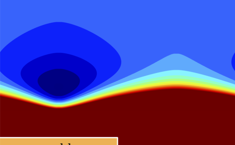
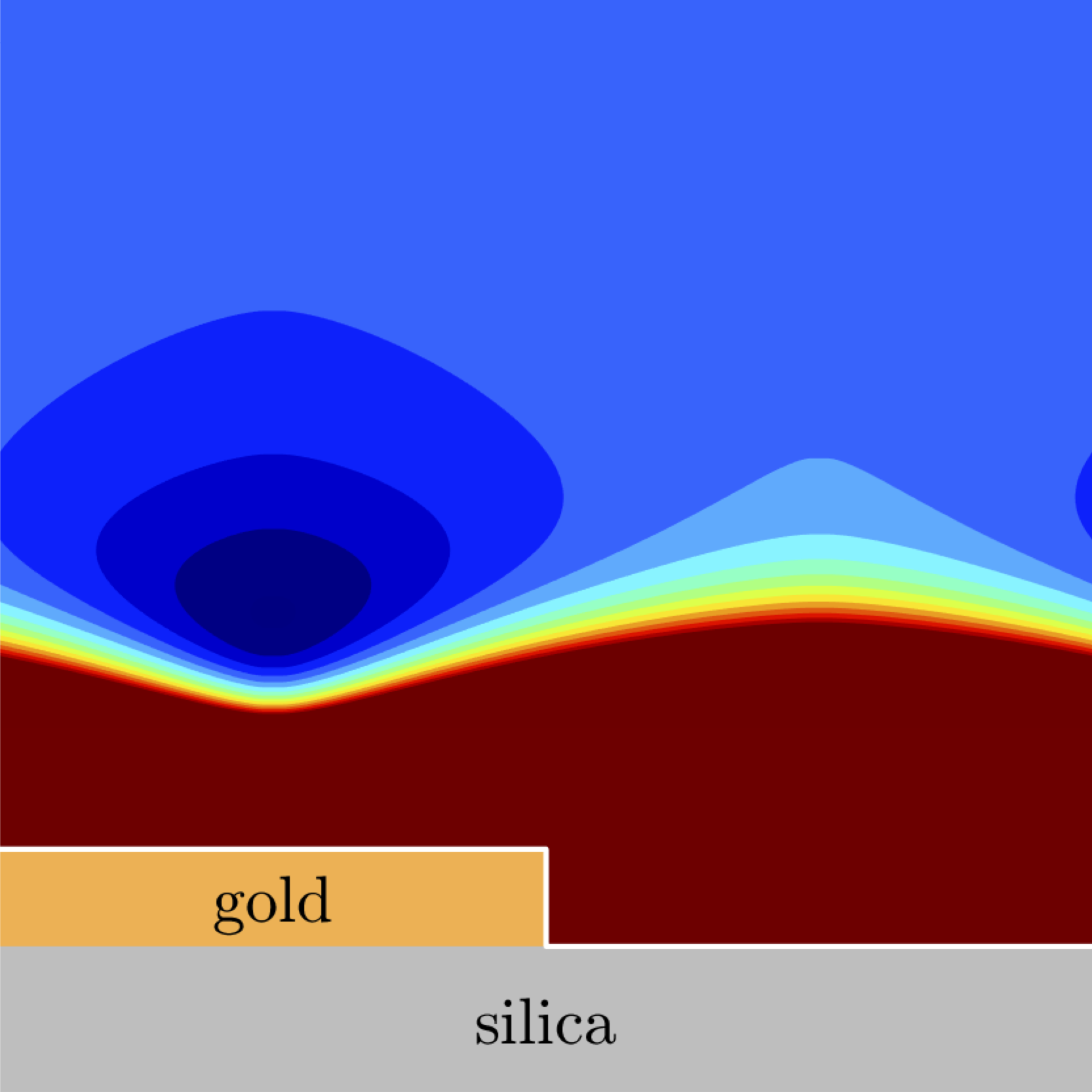
Experiments show that repulsive critical Casimir forces can be used to prevent stiction due to Casimir-Lifshitz forces. In a recent work, a microscopic metallic flake was suspended in a liquid solution above a metal-coated substrate [1]. By suspending the flake in a binary critical mixture and tuning the temperature we can control the flake’s hovering height above the substrate and, in the case of repulsive critical Casimir forces, prevent stiction.
Here, we present the model for the system of the metallic flake suspended above a metal-coated substrate in a binary critical mixture and show that repulsive critical Casimir forces can effectively counteract Casimir-Lifshitz forces and can be used to control dynamically the height of the flake above the surface. This provides a validation of the experimental results and a base to explore and design the behavior of similar systems in view of micro- and nanotechnological applications.
References
[1] F. Schmidt, A. Callegari, A. Daddi-Moussa-Ider, B. Munkhbat, R. Verre, T. Shegai, M. Käll, H. Löwen, A. Gambassi and G. Volpe, to be submitted (2022)