Poster by Sreekanth K Manikandan at the Protein Folding in Real Time conference, Stockholm, 11 March 2026

Recent advances in nonequilibrium physics allow extracting thermodynamic quantities, such as entropy production, directly from dynamical information in microscopic movies. (Figure by S. Manikandan, adapted from Manikandan et al., Phys. Rev. Research 6, 023310 (2024).)
Probing the Non-equilibrium Dynamics of Living Matter
Sreekanth K Manikandan
Date: 11 March 2026
Time: 18.00-20.00
Place: Aula Medica, Stockholm Sweden
Conference Protein Folding in Real Time, 11-13 March 2026, Stockholm, Sweden

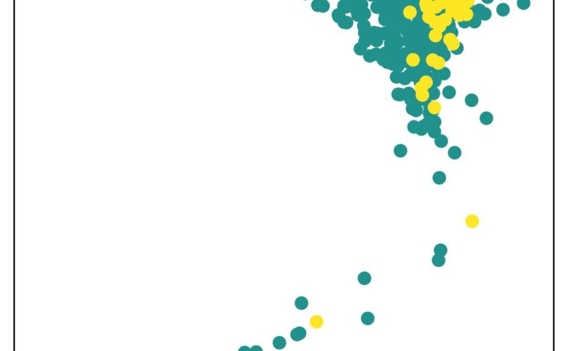
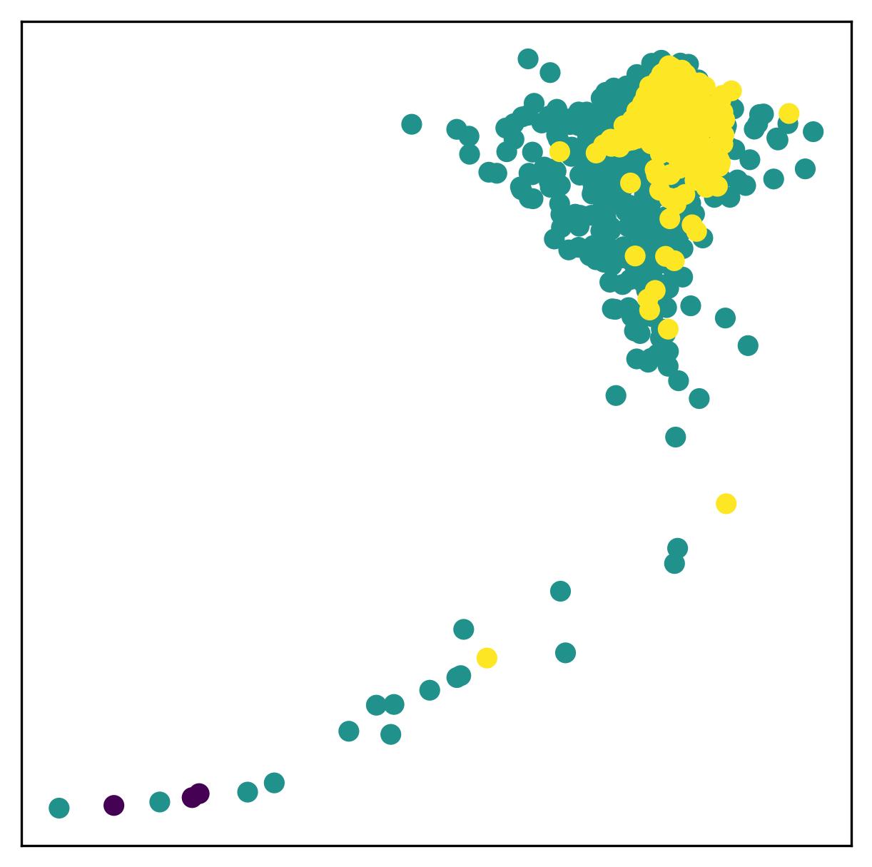
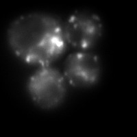
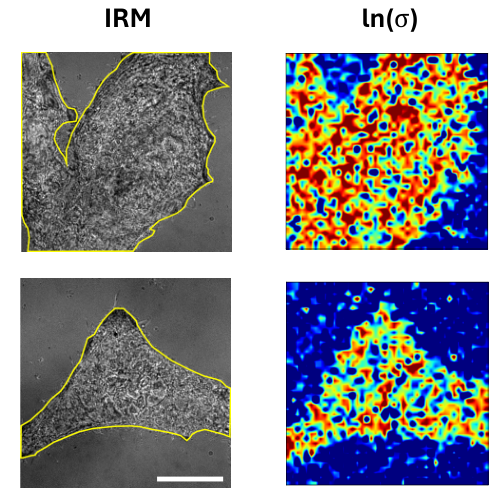
Identifying whether a process is in equilibrium, quantifying its distance from equilibrium, and constructing optimal reduced descriptions of non-equilibrium dynamics remain central challenges in the study of living matter. Here, we discuss how data-driven approaches grounded in stochastic thermodynamics enable these features to be inferred directly from experimental data. In particular, we show how entropy production can be localized in space and time, and how maximally dissipative coordinates emerge as effective low-dimensional descriptions of non-equilibrium processes. We highlight applications to experimental biophysical systems and discuss key challenges and limitations.

Poster by M. Selin at the Protein Folding in Real Time conference, Stockholm, 11 March 2026

Illustration of three different experiments autonomously performed by the SmartTrap system: DNA pulling experiments (top), red blood cell stretching (bottom left), and particle-particle interaction measurements (bottom right). (Image by M. Selin.)
SmartTrap: Automation of single molecule experiments
Martin Selin, Antonio Ciarlo, Giuseppe Pesce, Lars Bengtsson, Joan Camunas-Soler, Vinoth Sundar Rajan, Fredrik Westerlund, L. Marcus Wilhelmsson, Isabel Pastor, Felix Ritort, Steven B. Smith, Carlos Bustamante, and Giovanni Volpe
Date: 11th March 2026
Time: 18:00 – 20:00
Place: Aula Medica, Karolinska Institute, Solna
Conference Protein Folding in Real Time, 11-13 March 2026, Stockholm, Sweden

Single-molecule studies are vital for understanding fundamental biological processes, including protein folding, DNA transcription, and replication. However, performing these experiments manually on individual molecules is notoriously time-consuming and costly. To address this challenge, we have developed a fully autonomous single-molecule force spectroscopy platform by integrating a custom-built optical tweezers instrument with real-time deep-learning-based image analysis and adaptive control protocols. Our system achieves human-level throughput in terms of experiments per hour while remaining robust enough to operate continuously for hours without intervention. We demonstrate the versatility of our platform by having it perform DNA pulling experiments fully autonomously. By making the software open source we democratize high-throughput data collection in single-molecule biophysics, paving the way for merging single-molecule studies with large-scale, data-driven approaches—ultimately enabling new insights into the dynamic, transient states of complex biological systems.

Poster by A. Schiano di Colella at the Protein Folding in Real Time conference, Stockholm, 11 March 2026

Schematic representation of the architecture of a quantum circuit for application in variational problems. (Image by A. Schiano di Colella.)
Quantum computing for variational problems
Andrea Schiano di Colella, Antonio Ciarlo, Mats Granath, Giovanni Volpe
Date: 11 March 2026
Time: 18:00 – 20:00
Place: Aula Medica, Karolinska Institutet, Stockholm, Sweden
Conference: Protein Folding in Real Time

Quantum computing is a field of study that aims to exploit quantum mechanical effects for the purposes of computation. Due to the intrinsic capacity of qubits of efficiently represent an exponentially large configuration space, quantum computation has been identified as a promising candidate for complex physical chemistry simulations, including investigating the dynamics of protein folding. This work illustrates the use of quantum computing for variational problems, and the use of alternative training methods such as genetic algorithms to avoid the “barren plateau” phenomenon, which prevents the training of general quantum circuits by means of the usual gradient descent.

Poster by J. Dominguez at the Protein Folding in Real Time conference, Stockholm, 11 March 2026

Brightfield image of biofilm inside the droplet where motile aggregates are highlighted in yellow, biofilm mass in blue, and open patches in the structures due to dispersal in orange. (Image from D. Pérez and J. Domínguez)
Quantitative Analysis of Dynamic Biofilm Structures via Time-Resolved Droplet Microfluidics and Artificial Intelligence
Daniela Pérez Guerrero, Jesús Manuel Antúnez Domínguez, Aurélie Vigne, Daniel Midtvedt, Wylie Ahmed, Lisa Muiznieks, Giovanni Volpe and Caroline Beck Adiels
Date: 11th March 2026
Time: 18:00 – 20:00
Place: Aula Medica, Karolinska Institute, Solna
Conference Protein Folding in Real Time, 11-13 March 2026, Stockholm, Sweden

Droplet Microfluidics offers a powerful approach to study the spatiotemporal dynamics of biofilm formation at high resolution and throughput. By encapsulating microbial communities within controlled microenvironments, it becomes possible to monitor biofilm development continuously using time-lapse imaging, capturing transitions from initial attachment to maturation and dispersal. To make sense of these complex, high-dimensional datasets, we are developing an unsupervised variational autoencoder framework that can automatically identify and separate distinct stages of biofilm growth without prior labeling. This approach enables the extraction of latent features that characterize structural and behavioral shifts within the biofilm over time. In this context, protein folding may play a critical role in regulating both the establishment and dispersal of biofilms, as the conformational states of key structural and regulatory proteins can influence adhesion, matrix production, and the transition back to planktonic states.

Poster by J. Tember at the Protein Folding in Real Time conference, Stockholm, 11 March 2026

Comparison of anisotropic light-sheet microscopy data and super-resolved reconstruction, illustrating improved continuity and structural clarity of volumetric features. (Image by J. Tember.)
FIREFLY – Framework for Interactive Rendering, Exploration and Feature Extraction for Light-Sheet Microscopy
John Tember, Harshith Bachimanchi, Tsz Long Chu, Xin Tian, Andrei Chagin, Giovanni Volpe
Date: 11th March 2026
Time: 18:00 – 20:00
Place: Aula Medica, Karolinska Institute, Solna
Conference: Protein Folding in Real Time, 11-13 March 2026, Stockholm, Sweden

We present FIREFLY (Framework for Interactive Rendering, Exploration and Feature Extraction for Light-Sheet Microscopy), a platform for scalable computational tools. Modern 3D microscopy datasets are increasingly large and complex, posing challenges for efficient visualization, processing, and quantitative analysis. Our work focuses on developing tools and methods to support interactive and high-quality analysis workflows.

We are building a Unity-based application for real-time, multi-channel 3D rendering, enabling interactive inspection and quantitative analysis of large volumetric datasets. In parallel, we explore self-supervised machine learning approaches for enhancing anisotropic microscopy volumes and improving resolution without requiring additional training data.

Together, these efforts aim to provide an integrated and scalable pipeline for visualization and analysis in light-sheet microscopy.

Poster by N. C. Palmero Cruz at the Protein Folding in Real Time conference, Stockholm, 11 March 2026

3D model of the integrated setup combining optical tweezers, light-sheet microscopy, and microfluidics to manipulate the gut microbiome in vivo in zebrafish. (Image by N. C. Palmero Cruz.)
Optical Manipulation of Gut Microbiome and Neural Responses in Zebrafish
Norma Caridad Palmero Cruz
Date: 11 March 2026
Time: 18.00-20.00
Place: Aula Medica, Stockholm Sweden
Conference Protein Folding in Real Time, 11-13 March 2026, Stockholm, Sweden

The gut-brain axis is a complex, bidirectional network linking the microbiome to the central nervous system, significantly affecting physiological processes and neurological health, including conditions like autism and depression. Due to the genetic similarities between zebrafish and humans, the zebrafish serves as a valuable model for investigating the bidirectional relationship between the gut and brain, offering insights into how it compares with human behaviors. Research on the connection between gut and brain development typically involves using germ-free lab animals, where the microbiome is eliminated, and comparing them to those with restored microbiomes. However, this method does not capture the complexity of microbiome-nervous system communication due to its all-or-nothing approach. This work presents a setup that combines microfluidic techniques, optical tweezers, and light sheet microscopy to precisely manipulate the microbiome in larval zebrafish in situ and in vivo. This approach offers deeper insights into gut-brain connectivity and its impact on neurological health.

Poster by J. Huang at the Protein Folding in Real Time conference, Stockholm, 11 March 2026

The embedding space generated byvariational autoencoder (VAE), with samples colored by data quality. (Image by J. Huang)
Machine Learning-based Data Quality Control for AFM Force Spectroscopy
Jiacheng Huang, Nazli Demirpehlivan, Prakhar Dutta, Rahul Nagshi, Thomas Catley, Sylvia Whittle, Carlo Manzo, Rachel Owen, Giovanni Volpe
Date: 11th March 2026
Time: 18:00 – 20:00
Place: Aula Medica, Karolinska Institute, Solna
Conference Protein Folding in Real Time, 11-13 March 2026, Stockholm, Sweden

Atomic Force Microscopy (AFM) force spectroscopy is widely used to probe the mechanical properties and interactions of biological samples at the nanoscale, including living cells. However, large datasets generated during AFM measurements often contain curves affected by experimental artifacts such as poor tip–sample contact, noise, or instrumental instability. These low-quality force curves can significantly affect downstream analysis and typically require time-consuming manual inspection. In this work, we propose a machine learning–based data quality control framework for AFM force spectroscopy using a self-supervised approach.

Poster by L. Viaene at the Protein Folding in Real Time conference, Stockholm, 11 March 2026

Heat-induced aggregates in Saccharomyces cerevisiae on Slimfield SMLM. Hsp104-mGFP binds to misfolded regions enabling aggregate visualisation for tracking. (Image by L. Viaene.)
A single-molecule approach to study the spatial protein quality control system
Linde Viaene
Date: 11 March 2026
Time: 18.00-20.00
Place: Aula Medica, Stockholm Sweden
Conference Protein Folding in Real Time, 11-13 March 2026, Stockholm, Sweden

Cell populations are inherently diverse, and averaging measurements across them can mask subtle or rare cellular behaviours. In this work, we use live Slimfield single-molecule microscopy to study the role of Hsp104 in clearing misfolded and aggregated proteins after stress. By analysing endogenously tagged Hsp104, we quantify molecular diffusion and stoichiometry before and after heat stress. Our results show a transition from faster, more mobile molecules to larger, more static assemblies following stress, consistent with Hsp104 functionally engaging with protein aggregates. These measurements provide molecular-level insight into how cells respond to proteotoxic stress.

Invited talk by S. K. Manikandan at The 15th Nordic Workshop on Statistical Physics, Nordita, 26 Feb 2026

Recent advances in nonequilibrium physics allow extracting thermodynamic quantities, such as entropy production, directly from dynamical information in microscopic movies. (Figure by S. Manikandan, adapted from Manikandan et al., Phys. Rev. Research 6, 023310 (2024).)
Localising Entropy Production and Maximally Dissipative Coordinates from Experimental Data
Sreekanth Manikandan
Date: 26th February 2026
Time: 14.15
Place: NORDITA, Stockholm, Sweden
The 15th Nordic Workshop on Statistical Physics: Biological, Complex and Non-equilibrium Systems

Identifying whether a process is in equilibrium, quantifying how far it lies from equilibrium, and determining optimal reduced descriptions of non-equilibrium processes remain challenging open problems. Here, we discuss how novel data-driven techniques grounded in stochastic thermodynamics can be used to efficiently learn these features directly from experimental data. In particular, we show how entropy production can be localized in space and time, and how maximally dissipative coordinates can be consistently inferred as effective low-dimensional descriptions of non-equilibrium processes. We further discuss applications to experimental biophysical systems and outline key challenges and limitations.

Photos

Sreekanth, presenting. (Photo by A. Ciarlo)

Invited talk by A. Ciarlo at The 15th Nordic Workshop on Statistical Physics, Nordita, 26 Feb 2026

Schematic illustration of the light-momentum detection principle underlying SmartTrap. The momentum change of the trapping laser, induced by its interaction with the trapped particle, is measured to directly quantify optical forces with high precision, enabling real-time feedback and autonomous control in non-equilibrium experiments. (Figure by A. Ciarlo.)
SmartTrap: Autonomous Optical Tweezers for Statistical Physics of Non-Equilibrium Systems
Antonio Ciarlo
Date: 26th February 2026
Time: 13.30
Place: NORDITA, Stockholm, Sweden
The 15th Nordic Workshop on Statistical Physics: Biological, Complex and Non-equilibrium Systems

Optical tweezers are a key tool in non-equilibrium statistical physics, allowing direct measurements of forces, work, and fluctuations in single-molecule and soft matter systems. However, manual operation limits throughput and the systematic study of rare events.

In this talk, Antonio Ciarlo will present SmartTrap, a fully autonomous optical tweezers platform integrating deep learning–based 3D tracking, adaptive feedback control, and automated microfluidics. The system operates without human intervention, executing complete force spectroscopy protocols.

Demonstrated with high-throughput DNA pulling experiments on λ-DNA, SmartTrap enables precise measurements of force–extension curves and folding kinetics. The platform also opens new possibilities for studies of colloids, single cells, and quantitative tests of non-equilibrium statistical physics.

Photos

Antonio, presenting. (Photo by S. K. Manikandan)