Barbora Spackova joins the Soft Matter Lab at the Physics Department of the University of Gothenburg on 1st September 2021.
Barbora has a PhD in physical engineering from the Czech Technical University in Prague (Czech Republic). Formerly, she has been a researcher at Chalmers University of Technology in the group of Prof. Christoph Langhammer. Her research is focused on single-molecule detection in nanofluidic systems.
While part of the Soft Matter Lab, she will continue her research on characterising cell media containing exosomes using Nanofluidic Scattering Microscopy (NSM).
M. xanthus cell-cell and cell-particle local interactions during cellular aggregation.The environment topography alters the transition from single-cell populations to multicellular structures in Myxococcus xanthus
Karla C. Hernández Ramos, Edna Rodríguez-Sánchez, Juan Antonio Arias del Angel, Alejandro V. Arzola, Mariana Benítez, Ana E. Escalante, Alessio Franci, Giovanni Volpe, Natsuko Rivera-Yoshida
Sci. Adv. 7(35), eabh2278 (2021)
bioRxiv: 10.1101/2021.01.27.428527
doi: 10.1126/sciadv.abh2278
The social soil-dwelling bacteria Myxococcus xanthus can form multicellular structures, known as fruiting bodies. Experiments in homogeneous environments have shown that this process is affected by the physico-chemical properties of the substrate, but they have largely neglected the role of complex topographies. We experimentally demonstrate that the topography alters single-cell motility and multicellular organization in M. xanthus. In topographies realized by randomly placing silica particles over agar plates, we observe that the cells’ interaction with particles drastically modifies the dynamics of cellular aggregation, leading to changes in the number, size and shape of the fruiting bodies, and even to arresting their formation in certain conditions. We further explore this type of cell-particle interaction in a minimal computational model. These results provide fundamental insights into how the environment topography influences the emergence of complex multicellular structures from single cells, which is a fundamental problem of biological, ecological and medical relevance.
Age-independent cognitive connectome in the whole cohort.The Cognitive Connectome in Healthy Aging
Eloy Garcia-Cabello, Lissett Gonzalez-Burgos, Joana B. Pereira, Juan Andres Hernández-Cabrera, Eric Westman, Giovanni Volpe, José Barroso, & Daniel Ferreira
Front. Aging Neurosci. 13, 530 (2021)
doi: 10.3389/fnagi.2021.694254
Objectives: Cognitive aging has been extensively investigated using both univariate and multivariate analyses. Sophisticated multivariate approaches such as graph theory could potentially capture unknown complex associations between multiple cognitive variables. The aim of this study was to assess whether cognition is organized into a structure that could be called the “cognitive connectome,” and whether such connectome differs between age groups.
Methods: A total of 334 cognitively unimpaired individuals were stratified into early-middle-age (37–50 years, n = 110), late-middle-age (51–64 years, n = 106), and elderly (65–78 years, n = 118) groups. We built cognitive networks from 47 cognitive variables for each age group using graph theory and compared the groups using different global and nodal graph measures.
Results: We identified a cognitive connectome characterized by five modules: verbal memory, visual memory—visuospatial abilities, procedural memory, executive—premotor functions, and processing speed. The elderly group showed reduced transitivity and average strength as well as increased global efficiency compared with the early-middle-age group. The late-middle-age group showed reduced global and local efficiency and modularity compared with the early-middle-age group. Nodal analyses showed the important role of executive functions and processing speed in explaining the differences between age groups.
Conclusions: We identified a cognitive connectome that is rather stable during aging in cognitively healthy individuals, with the observed differences highlighting the important role of executive functions and processing speed. We translated the connectome concept from the neuroimaging field to cognitive data, demonstrating its potential to advance our understanding of the complexity of cognitive aging.
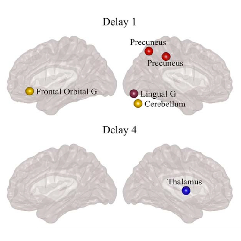
Differences between controls and PD participants in nodal network measures. (Image taken from the article.)Directed Brain Connectivity Identifies Widespread Functional Network Abnormalities in Parkinson’s Disease
Mite Mijalkov, Giovanni Volpe, Joana B Pereira
Cerebral Cortex, bhab237 (2021)
doi: 10.1093/cercor/bhab237
Parkinson’s disease (PD) is a neurodegenerative disorder characterized by topological abnormalities in large-scale functional brain networks, which are commonly analyzed using undirected correlations in the activation signals between brain regions. This approach assumes simultaneous activation of brain regions, despite previous evidence showing that brain activation entails causality, with signals being typically generated in one region and then propagated to other ones. To address this limitation, here, we developed a new method to assess whole-brain directed functional connectivity in participants with PD and healthy controls using antisymmetric delayed correlations, which capture better this underlying causality. Our results show that whole-brain directed connectivity, computed on functional magnetic resonance imaging data, identifies widespread differences in the functional networks of PD participants compared with controls, in contrast to undirected methods. These differences are characterized by increased global efficiency, clustering, and transitivity combined with lower modularity. Moreover, directed connectivity patterns in the precuneus, thalamus, and cerebellum were associated with motor, executive, and memory deficits in PD participants. Altogether, these findings suggest that directional brain connectivity is more sensitive to functional network differences occurring in PD compared with standard methods, opening new opportunities for brain connectivity analysis and development of new markers to track PD progression.
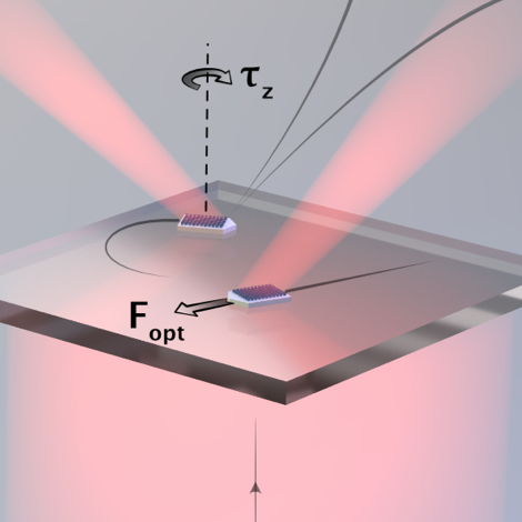
Metavehicles.Microscopic Metavehicles Powered and Steered by Embedded Optical Metasurfaces
Daniel Andrén, Denis G. Baranov, Steven Jones, Giovanni Volpe, Ruggero Verre, Mikael Käll
Nat. Nanotechnol. (2021)
doi: 10.1038/s41565-021-00941-0
arXiv: 2012.10205
Nanostructured dielectric metasurfaces offer unprecedented opportunities to manipulate light by imprinting an arbitrary phase gradient on an impinging wavefront. This has resulted in the realization of a range of flat analogues to classical optical components, such as lenses, waveplates and axicons. However, the change in linear and angular optical momentum associated with phase manipulation also results in previously unexploited forces and torques that act on the metasurface itself. Here we show that these optomechanical effects can be utilized to construct optical metavehicles – microscopic particles that can travel long distances under low-intensity plane-wave illumination while being steered by the polarization of the incident light. We demonstrate movement in complex patterns, self-correcting motion and an application as transport vehicles for microscopic cargoes, which include unicellular organisms. The abundance of possible optical metasurfaces attests to the prospect of developing a wide variety of metavehicles with specialized functional behaviours.
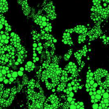
Virtually-stained generated image for lipid-droplet.Extracting quantitative biological information from bright-field cell images using deep learning
Saga Helgadottir, Benjamin Midtvedt, Jesús Pineda, Alan Sabirsh, Caroline B. Adiels, Stefano Romeo, Daniel Midtvedt, Giovanni Volpe
Biophysics Rev. 2, 031401 (2021)
arXiv: 2012.12986
doi: 10.1063/5.0044782
Quantitative analysis of cell structures is essential for biomedical and pharmaceutical research. The standard imaging approach relies on fluorescence microscopy, where cell structures of interest are labeled by chemical staining techniques. However, these techniques are often invasive and sometimes even toxic to the cells, in addition to being time-consuming, labor-intensive, and expensive. Here, we introduce an alternative deep-learning-powered approach based on the analysis of bright-field images by a conditional generative adversarial neural network (cGAN). We show that this approach can extract information from the bright-field images to generate virtually-stained images, which can be used in subsequent downstream quantitative analyses of cell structures. Specifically, we train a cGAN to virtually stain lipid droplets, cytoplasm, and nuclei using bright-field images of human stem-cell-derived fat cells (adipocytes), which are of particular interest for nanomedicine and vaccine development. Subsequently, we use these virtually-stained images to extract quantitative measures about these cell structures. Generating virtually-stained fluorescence images is less invasive, less expensive, and more reproducible than standard chemical staining; furthermore, it frees up the fluorescence microscopy channels for other analytical probes, thus increasing the amount of information that can be extracted from each cell.
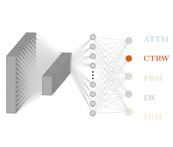
RANDI architecture to classify the model underlying anomalous diffusion.Classification, inference and segmentation of anomalous diffusion with recurrent neural networks
Aykut Argun, Giovanni Volpe, Stefano Bo
J. Phys. A: Math. Theor. 54 294003 (2021)
doi: 10.1088/1751-8121/ac070a
arXiv: 2104.00553
Countless systems in biology, physics, and finance undergo diffusive dynamics. Many of these systems, including biomolecules inside cells, active matter systems and foraging animals, exhibit anomalous dynamics where the growth of the mean squared displacement with time follows a power law with an exponent that deviates from 1. When studying time series recording the evolution of these systems, it is crucial to precisely measure the anomalous exponent and confidently identify the mechanisms responsible for anomalous diffusion. These tasks can be overwhelmingly difficult when only few short trajectories are available, a situation that is common in the study of non-equilibrium and living systems. Here, we present a data-driven method to analyze single anomalous diffusion trajectories employing recurrent neural networks, which we name RANDI. We show that our method can successfully infer the anomalous exponent, identify the type of anomalous diffusion process, and segment the trajectories of systems switching between different behaviors. We benchmark our performance against the state-of-the art techniques for the study of single short trajectories that participated in the Anomalous Diffusion (AnDi) challenge. Our method proved to be the most versatile method, being the only one to consistently rank in the top 3 for all tasks proposed in the AnDi challenge.
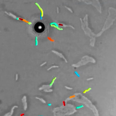
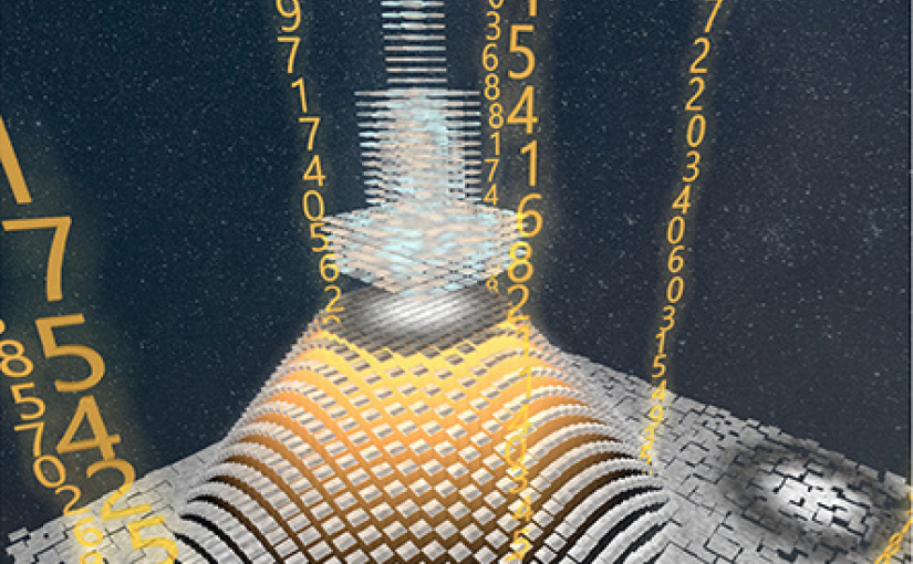
Deep learning for particle tracking. (Image by Aykut Argun)Deep learning for microscopy, optical trapping, and active matter
Giovanni Volpe
Colloquium
(online at) TU-Darmstadt, Germany
18 June 2021, 14:00 CEST
After a brief overview of artificial intelligence, machine learning and deep learning, I will present a series of recent works in which we have employed deep learning for applications in photonics and active matter.
In particular, I will explain how we employed deep learning to enhance digital video microscopy, to estimate the properties of anomalous diffusion, to characterize microscopic force fields, to improve the calculation of optical forces, and to characterize nanoparticles.
Finally, I will provide an outlook for the application of deep learning in photonics and active matter.
Olle Fager defended his Master thesis in MPCAS at the Chalmers University of Technology on 15 June 2021. Congrats!
Title: Real-Time Multi-Object Tracking and Segmentation with Generated Data using 3D-modelling
Multi-Object Tracking and Segmentation (MOTS) is an important branch of computer vision that has applications in many different areas. In recent developments these methods have been able to reach favorable speed-accuracy trade-offs, making them interesting for real-time applications. In this work different deep learning based MOTS methods have been investigated with the purpose of extending the DeepTrack framework with real-time MOTS capabilities. Deep learning methods rely heavily on the data on which they are trained. The collection and annotation of the data can however be very time-consuming. Therefor, a pipeline is developed and investigated that automatically produces synthetic data by utilizing 3D-modelling. The most accurate tracker achieves a MOTSA score of 94 and the tracker with the best speed-accuracy trade-off achieves a MOTSA score of 88. It is also observed that satisfactory results can be achieved in most situations with a quite general data generation pipeline, indicating that the developed pipeline could be used in different scenarios.
Name of the master programme: MPCAS – Complex Adaptive Systems Supervisor: Giovanni Volpe Examiner: Giovanni Volpe, Department of Physics, University of Gothenburg Opponent: Arianit Zeqiri and Morad Mahmoudyan