Presentation by B. García Rodríguez at SPIE-ETAI, San Diego, 5 August 2025

Biomolecular condensates are analyzed and characterized using a holographic microscope, which utilizes a variational autoencoder (VAE) to quantify size, protein concentration, and the sharpness of the RNA-binding protein interface. (Image by B. Garcia.)
Structure and dynamics of biomolecular condensates revealed by deep learning enhanced interferometric microscopy
Berenice García Rodríguez, Makasewicz Katarzyna, Giovanni Volpe, Paolo Arosio, Daniel Sundås Midtvedt.
Date: 5 August 2025
Time: 5:00 PM – 5:15 PM PDT
Place:
Conv. Ctr. Room 4
Presentation type:
Oral

Biomolecular condensates are biological structures that form through weak, multivalent interactions primarily between low complexity domains of intrinsically disordered proteins, existing in cells as submicrometer structures. Their functions rely sensitively on physical properties such as size, internal protein concentration, and interfacial tension. However, direct measurements of these properties in submicrometer condensates remain scarce. In this work, we employ deep learning enhanced interferometric imaging to quantify size, protein concentration, and the sharpness of the interface of submicrometer condensates formed by the low complexity domain of the RNA-binding protein DDX4-LCD. We find that, within the two-phase region, DDX4-LCD forms spherical condensates with an internal protein concentration that can be slightly modulated by adding salt to the solution. Furthermore, we find that multiple populations of protein clusters coexist in the sample, some persisting even outside the two-phase region of the phase diagram, separable by their interface properties and dynamics. This hints at a more complex phase diagram of DDX4-LCD condensation than previously anticipated.


Optical Label-Free Microscopy Characterization of Dielectric Nanoparticles published in Nanoscale

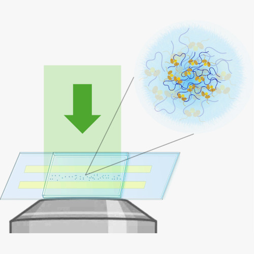
Propagation of scattered light through a scattering microscope, illustrating typical nanoparticles studied. (Image by B. García Rodriguez.)
Optical Label-Free Microscopy Characterization of Dielectric Nanoparticles
Berenice Garcia Rodriguez, Erik Olsén, Fredrik Skärberg, Giovanni Volpe, Fredrik Höök, Daniel Sundås Midtvedt
Nanoscale, 17, 8336-8362 (2025)
arXiv: 2409.11810
doi: 10.1039/D4NR03860F

In order to relate nanoparticle properties to function, fast and detailed particle characterization, is needed. The ability to characterize nanoparticle samples using optical microscopy techniques has drastically improved over the past few decades; consequently, there are now numerous microscopy methods available for detailed characterization of particles with nanometric size. However, there is currently no “one size fits all” solution to the problem of nanoparticle characterization. Instead, since the available techniques have different detection limits and deliver related but different quantitative information, the measurement and analysis approaches need to be selected and adapted for the sample at hand. In this tutorial, we review the optical theory of single particle scattering and how it relates to the differences and similarities in the quantitative particle information obtained from commonly used microscopy techniques, with an emphasis on nanometric (submicron) sized dielectric particles. Particular emphasis is placed on how the optical signal relates to mass, size, structure, and material properties of the detected particles and to its combination with diffusivity-based particle sizing. We also discuss emerging opportunities in the wake of new technology development, with the ambition to guide the choice of measurement strategy based on various challenges related to different types of nanoparticle samples and associated analytical demands.