Latent Space-Driven Quantification of Biofilm Formation using Time Resolved Droplet Microfluidics on ArXiv

Automated segnmentation of bacterial structures within a droplet. The image shows a bright-field microscopy view where a large biofilm region (green, outlined in blue) has been segmented from surrounding features. Small aggregates (yellow contours) are also highlighted. This segmentation enables structural differentiation of biofilm components for downstream quantitative analysis. (Image by D. Pérez Guerrero.)
Latent Space-Driven Quantification of Biofilm Formation using Time Resolved Droplet Microfluidics
Daniela Pérez Guerrero, Jesús Manuel Antúnez Domínguez, Aurélie Vigne, Daniel Midtvedt, Wylie Ahmed, Lisa D. Muiznieks, Giovanni Volpe, Caroline Beck Adiels
arXiv: 2507.07632

Bacterial biofilms play a significant role in various fields that impact our daily lives, from detrimental public health hazards to beneficial applications in bioremediation, biodegradation, and wastewater treatment. However, high-resolution tools for studying their dynamic responses to environmental changes and collective cellular behavior remain scarce. To characterize and quantify biofilm development, we present a droplet-based microfluidic platform combined with an image analysis tool for in-situ studies. In this setup, Bacillus subtilis was inoculated in liquid Lysogeny Broth microdroplets, and biofilm formation was examined within emulsions at the water-oil interface. Bacteria were encapsulated in droplets, which were then trapped in compartments, allowing continuous optical access throughout biofilm formation. Droplets, each forming a distinct microenvironment, were generated at high throughput using flow-controlled pressure pumps, ensuring monodispersity. A microfluidic multi-injection valve enabled rapid switching of encapsulation conditions without disrupting droplet generation, allowing side-by-side comparison. Our platform supports fluorescence microscopy imaging and quantitative analysis of droplet content, along with time-lapse bright-field microscopy for dynamic observations. To process high-throughput, complex data, we integrated an automated, unsupervised image analysis tool based on a Variational Autoencoder (VAE). This AI-driven approach efficiently captured biofilm structures in a latent space, enabling detailed pattern recognition and analysis. Our results demonstrate the accurate detection and quantification of biofilms using thresholding and masking applied to latent space representations, enabling the precise measurement of biofilm and aggregate areas.
Seminar by W. Ahmed on 13 March 2024

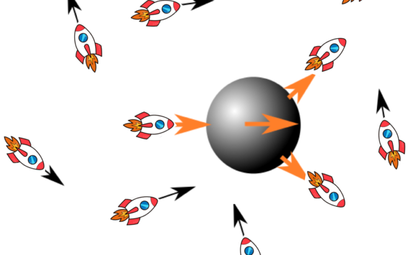
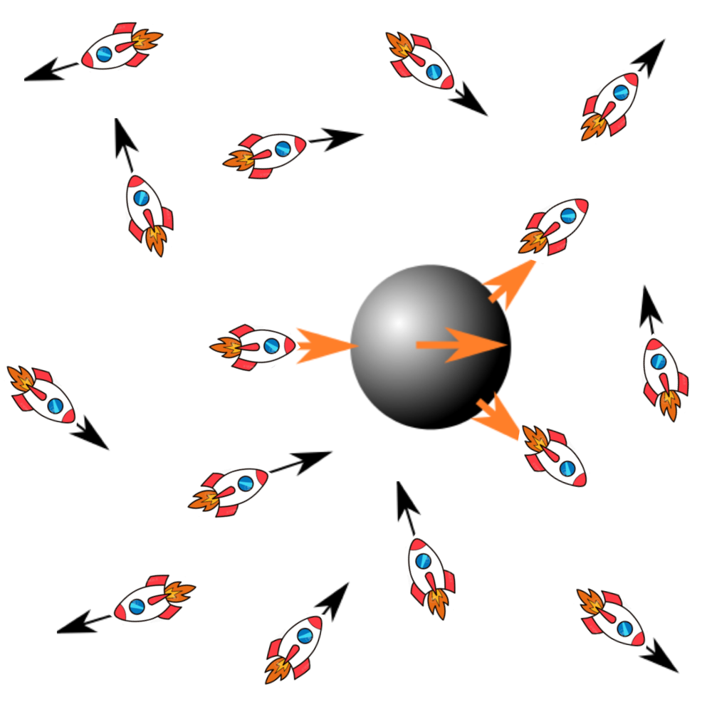
A schematic of a passive particle immersed in an active bath experiencing non-equilibrium fluctuations. (Illustration by W. Ahmed)
Emergent behavior in active biological matter
Wylie Ahmed
Laboratoire de Physique Theorique, Toulouse (France) and California State University, Fullerton (USA)

13 March 2024, 12:30, Nexus

Motivated by nucleus centering in mouse oocytes, we explore a different type of biological active matter. We investigate the stochastic force fluctuations of micro swimmers in two scenarios: (1) a single swimmer navigating through a passive fluid; (2) a dense suspension of swimmers surrounding a passive tracer. By direct force measurement using optical tweezers we show that the force trajectory of an individual micro swimmer exhibits rich oscillatory dynamics that vary in time. Interestingly, when these highly fluctuating force dynamics are analyzed using the framework of stochastic thermodynamics we recover energy dissipation rates in agreement with time-averaged fluid dynamics studies. For a dense suspension of swimmers serving as an active bath for a passive tracer we observe both shear thinning and thickening, which depends on Peclet number, and enhanced diffusion of our tracer by a factor of 2. We estimate the energy transfer rate from the active bath to the passive tracer. These two scenarios allow us to explore energy exchange between an active swimmer in a passive bath and a passive tracer in an active bath.

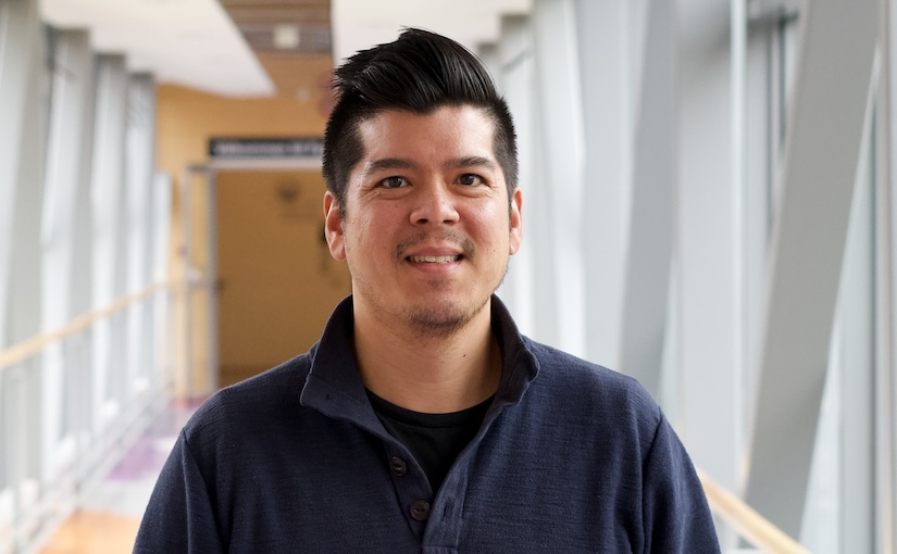
Wylie Ahmed visits the Soft Matter Lab. Welcome!

(Photo by A. Ciarlo)
Wylie Ahmed is a Visiting Professor from the Laboratoire de Physique Theorique in Toulouse, France. He is also an associate professor (on leave) at California State University, Fullerton where he leads the Laboratory for Soft, Living, and Active Matter (SLAMLab). His visiting position is financed through the CNRS with partial support from the Soft Matter Lab.
He will visit us for 5 months from March 1, 2024, to July 31, 2024.

He completed his Ph.D. at the University of Illinois at Urbana-Champaign, and was a Marie Skłodowska-Curie Research Fellow at the Institut Curie in Paris, France. He started his group in 2016 in California and is now moving his research activities to Toulouse France. His research interests are in cellular biophysics, soft and active matter physics, and bio-inspired materials with a theme towards understanding emergent behavior.