After a brief overview of artificial intelligence, machine learning and deep learning, I will present a series of recent works in which we have employed deep learning for applications in photonics and active matter. In particular, I will explain how we employed deep learning to enhance digital video microscopy, to estimate the properties of anomalous diffusion, to characterize microscopic force fields, to improve the calculation of optical forces, and to characterize nanoparticles. Finally, I will provide an outlook for the application of deep learning in photonics and active matter.
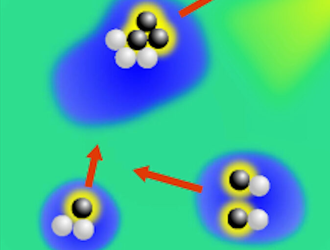
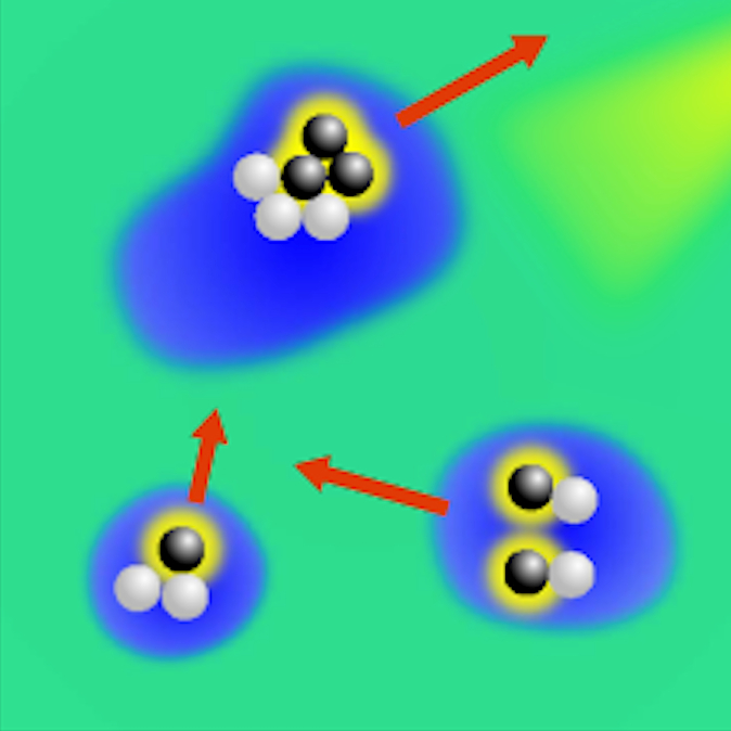
Active droploids. (Image taken from the article.)Active droploids
Jens Grauer, Falko Schmidt, Jesús Pineda, Benjamin Midtvedt, Hartmut Löwen, Giovanni Volpe & Benno Liebchen
Nat. Commun. 12, 6005 (2021)
doi: 10.1038/s41467-021-26319-3
arXiv: 2109.10677
Active matter comprises self-driven units, such as bacteria and synthetic microswimmers, that can spontaneously form complex patterns and assemble into functional microdevices. These processes are possible thanks to the out-of-equilibrium nature of active-matter systems, fueled by a one-way free-energy flow from the environment into the system. Here, we take the next step in the evolution of active matter by realizing a two-way coupling between active particles and their environment, where active particles act back on the environment giving rise to the formation of superstructures. In experiments and simulations we observe that, under light-illumination, colloidal particles and their near-critical environment create mutually-coupled co-evolving structures. These structures unify in the form of active superstructures featuring a droplet shape and a colloidal engine inducing self-propulsion. We call them active droploids—a portmanteau of droplet and colloids. Our results provide a pathway to create active superstructures through environmental feedback.
The study, recently published in Biophysics Reviews, shows how artificial intelligence can be used to develop faster, cheaper and more reliable information about cells, while also eliminating the disadvantages from using chemicals in the process.
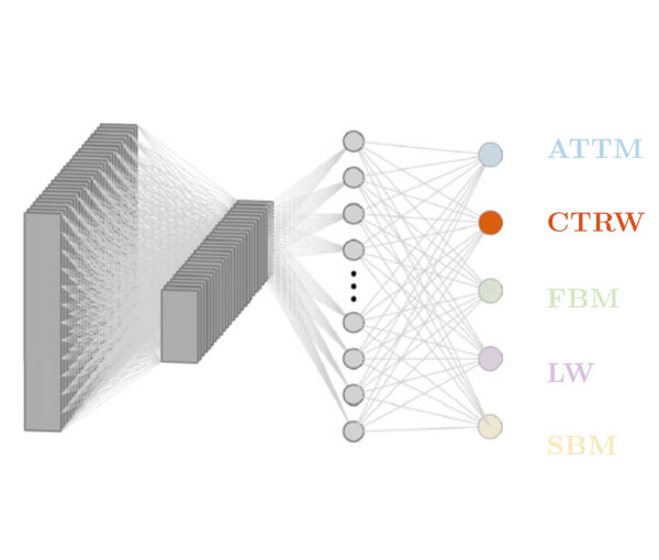
Countless systems in biology, physics, and finance undergo diffusive dynamics. Many of these systems, including biomolecules inside cells, active matter systems and foraging animals, exhibit anomalous dynamics where the growth of the mean squared displacement with time follows a power law with an exponent that deviates from 1. When studying time series recording the evolution of these systems, it is crucial to precisely measure the anomalous exponent and confidently identify the mechanisms responsible for anomalous diffusion. These tasks can be overwhelmingly difficult when only few short trajectories are available, a situation that is common in the study of non-equilibrium and living systems. Here, we present a data-driven method to analyze single anomalous diffusion trajectories employing recurrent neural networks, which we name RANDI. We show that our method can successfully infer the anomalous exponent, identify the type of anomalous diffusion process, and segment the trajectories of systems switching between different behaviors. We benchmark our performance against the state-of-the art techniques for the study of single short trajectories that participated in the Anomalous Diffusion (AnDi) challenge. Our method proved to be the most versatile method, being the only one to consistently rank in the top 3 for all tasks proposed in the AnDi challenge.
DeepTrack 2.0 Logo. (Image from DeepTrack 2.0 Project)Quantitative Digital Microscopy with Deep Learning Giovanni Volpe
Invited Talk at the Virtual school “Machine Learning and Automated Experiment in Scanning Probe Microscopy”
Online
October 4-7, 2021
11:20 AM
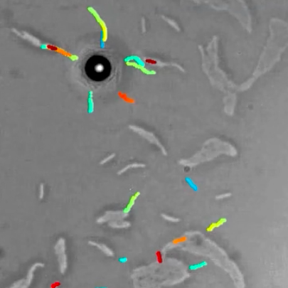
Video microscopy has a long history of providing insights and breakthroughs for a broad range of disciplines, from physics to biology. Image analysis to extract quantitative information from video microscopy data has traditionally relied on algorithmic approaches, which are often difficult to implement, time consuming, and computationally expensive. Recently, alternative data-driven approaches using deep learning have greatly improved quantitative digital microscopy, potentially offering automatized, accurate, and fast image analysis. However, the combination of deep learning and video microscopy remains underutilized primarily due to the steep learning curve involved in developing custom deep-learning solutions. To overcome this issue, we introduce a software, DeepTrack 2.0, to design, train and validate deep- learning solutions for digital microscopy. We use it to exemplify how deep learning can be employed for a broad range of applications, from particle localization, tracking and characterization to cell counting and classification. Thanks to its user- friendly graphical interface, DeepTrack 2.0 can be easily customized for user-specific applications, and, thanks to its open-source object-oriented programming, it can be easily expanded to add features and functionalities, potentially introducing deep-learning-enhanced video microscopy to a far wider audience.
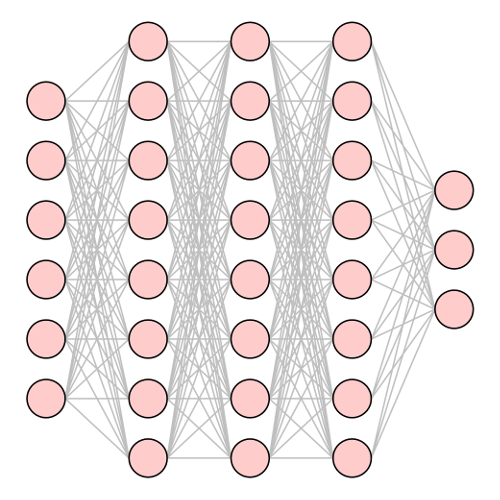
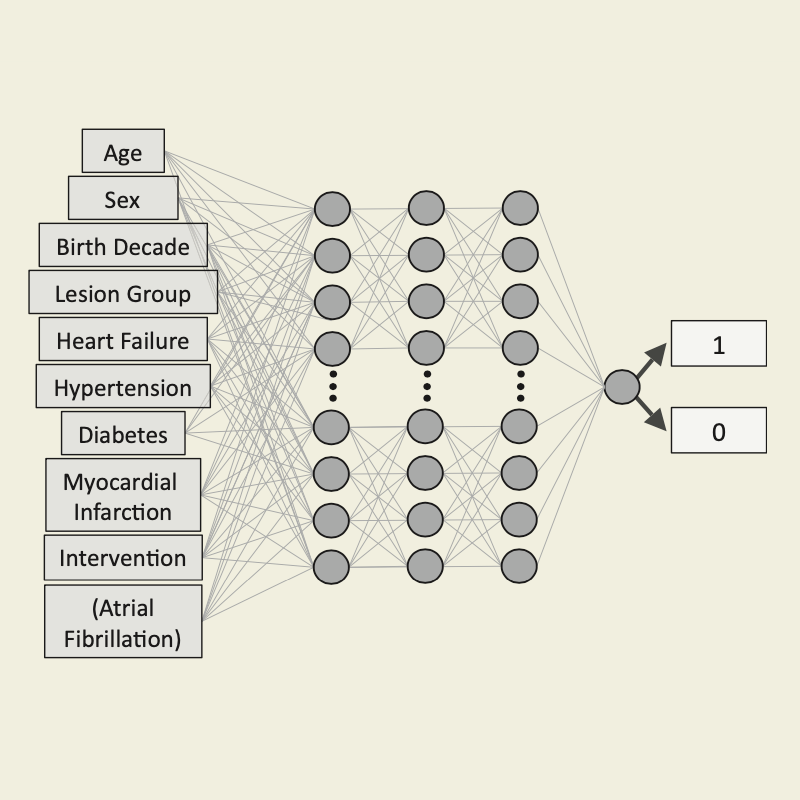
Neural net with input layer (left), dense internal layers, and output layer (right). (Image from the article Machine Learning for Active Matter)Machine Learning for Active Matter: Opportunities and Challenges
Giovanni Volpe
Invited Talk
(online at) ICTP, Trieste, Italy
8 September 2021, 11:30 CEST
Machine-learning methods are starting to shape active-matter research. Which new trends will this start? Which new groundbreaking insight and applications can we expect? More fundamentally, what can this contribute to our understanding of active matter? Can this help us to identify unifying principles and systematise active matter? This presentation addresses some of these questions with some concrete examples, exploring how machine learning is steering active matter towards new directions, offering unprecedented opportunities and posing practical and fundamental challenges. I will illustrate some most successful recent applications of machine learning to active matter with a slight bias towards work done in my research group: enhancing data acquisition and analysis; providing new data-driven models; improving navigation and search strategies; offering insight into the emergent dynamics of active matter in crowded and complex environments. I will discuss the opportunities and challenges that are emerging: implementing feedback control; uncovering underlying principles to systematise active matter; understanding the behaviour, organisation and evolution of biological active matter; realising active matter with embodied intelligence. Finally, I will highlight how active matter and machine learning can work together for mutual benefit.
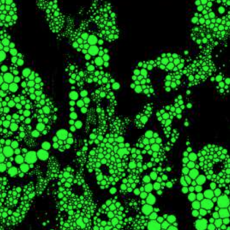
M. xanthus cell-cell and cell-particle local interactions during cellular aggregation.The environment topography alters the transition from single-cell populations to multicellular structures in Myxococcus xanthus
Karla C. Hernández Ramos, Edna Rodríguez-Sánchez, Juan Antonio Arias del Angel, Alejandro V. Arzola, Mariana Benítez, Ana E. Escalante, Alessio Franci, Giovanni Volpe, Natsuko Rivera-Yoshida
Sci. Adv. 7(35), eabh2278 (2021)
bioRxiv: 10.1101/2021.01.27.428527
doi: 10.1126/sciadv.abh2278
The social soil-dwelling bacteria Myxococcus xanthus can form multicellular structures, known as fruiting bodies. Experiments in homogeneous environments have shown that this process is affected by the physico-chemical properties of the substrate, but they have largely neglected the role of complex topographies. We experimentally demonstrate that the topography alters single-cell motility and multicellular organization in M. xanthus. In topographies realized by randomly placing silica particles over agar plates, we observe that the cells’ interaction with particles drastically modifies the dynamics of cellular aggregation, leading to changes in the number, size and shape of the fruiting bodies, and even to arresting their formation in certain conditions. We further explore this type of cell-particle interaction in a minimal computational model. These results provide fundamental insights into how the environment topography influences the emergence of complex multicellular structures from single cells, which is a fundamental problem of biological, ecological and medical relevance.
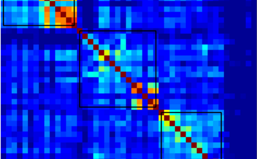
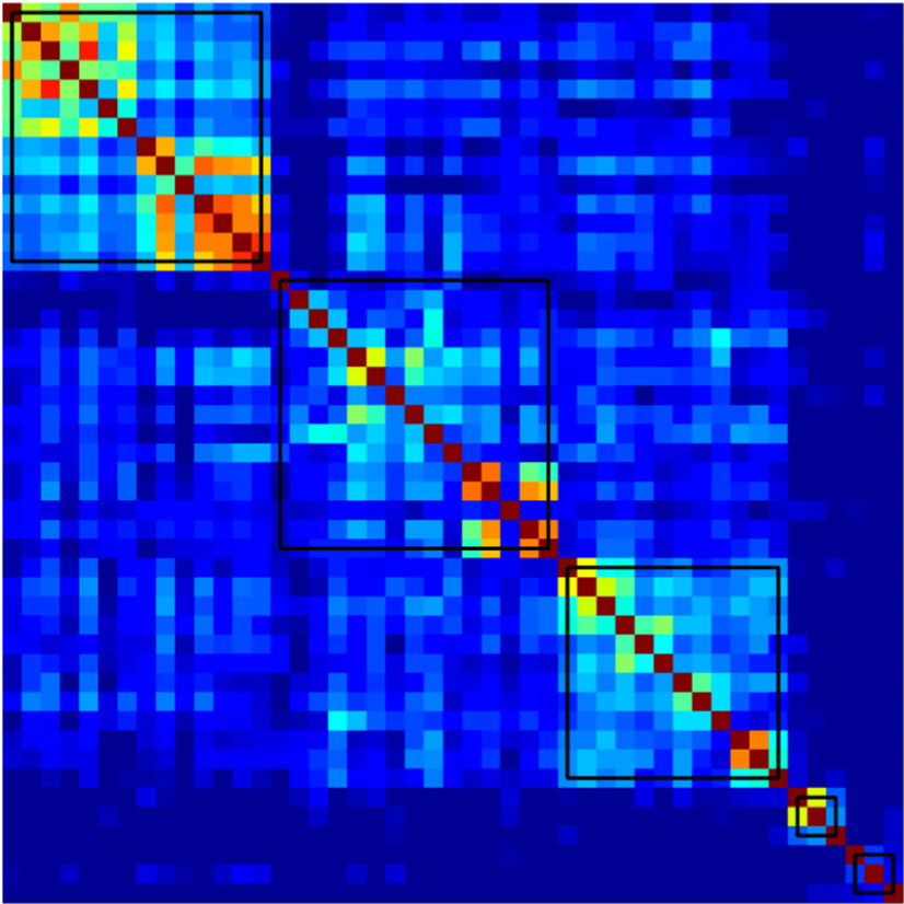
Age-independent cognitive connectome in the whole cohort.The Cognitive Connectome in Healthy Aging
Eloy Garcia-Cabello, Lissett Gonzalez-Burgos, Joana B. Pereira, Juan Andres Hernández-Cabrera, Eric Westman, Giovanni Volpe, José Barroso, & Daniel Ferreira
Front. Aging Neurosci. 13, 530 (2021)
doi: 10.3389/fnagi.2021.694254
Objectives: Cognitive aging has been extensively investigated using both univariate and multivariate analyses. Sophisticated multivariate approaches such as graph theory could potentially capture unknown complex associations between multiple cognitive variables. The aim of this study was to assess whether cognition is organized into a structure that could be called the “cognitive connectome,” and whether such connectome differs between age groups.
Methods: A total of 334 cognitively unimpaired individuals were stratified into early-middle-age (37–50 years, n = 110), late-middle-age (51–64 years, n = 106), and elderly (65–78 years, n = 118) groups. We built cognitive networks from 47 cognitive variables for each age group using graph theory and compared the groups using different global and nodal graph measures.
Results: We identified a cognitive connectome characterized by five modules: verbal memory, visual memory—visuospatial abilities, procedural memory, executive—premotor functions, and processing speed. The elderly group showed reduced transitivity and average strength as well as increased global efficiency compared with the early-middle-age group. The late-middle-age group showed reduced global and local efficiency and modularity compared with the early-middle-age group. Nodal analyses showed the important role of executive functions and processing speed in explaining the differences between age groups.
Conclusions: We identified a cognitive connectome that is rather stable during aging in cognitively healthy individuals, with the observed differences highlighting the important role of executive functions and processing speed. We translated the connectome concept from the neuroimaging field to cognitive data, demonstrating its potential to advance our understanding of the complexity of cognitive aging.
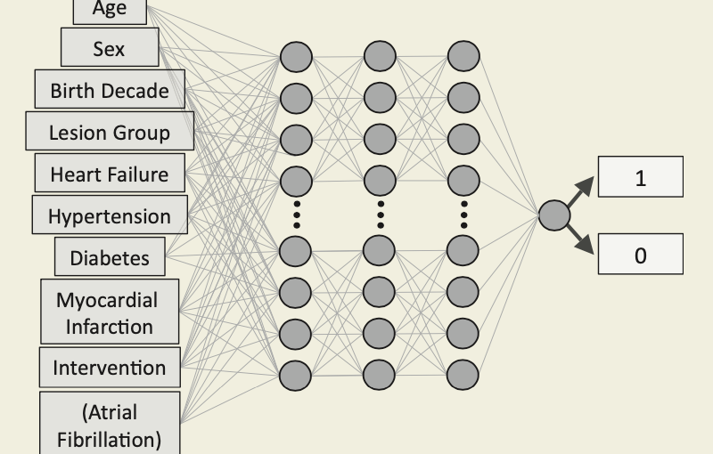
Neural network prediction of mortality and atrial fibrillation. (Image taken from the article’s graphical abstract.)Enhanced prediction of atrial fibrillation and mortality among patients with congenital heart disease using nationwide register based medical hospital data and neural networks
Kok Wai Giang, Saga Helgadottir, Mikael Dellborg, Giovanni Volpe, Zacharias Mandalenakis
European Heart Journal – Digital Health (2021)
doi: 10.1093/ehjdh/ztab065
Aims: To improve short-and long-term predictions of mortality and atrial fibrillation (AF) among patients with congenital heart disease (CHD) from a nationwide population using neural networks (NN).
Methods and results: The Swedish National Patient Register and the Cause of Death Register were used to identify all patients with CHD born from 1970 to 2017. A total of 71 941 CHD patients were identified and followed-up from birth until the event or end of study in 2017. Based on data from a nationwide population, a NN model was obtained to predict mortality and AF. Logistic regression (LR) based on the same data was used as a baseline comparison. Of 71 941 CHD patients, a total of 5768 died (8.02%) and 995 (1.38%) developed AF over time with a mean follow-up time of 16.47 years (standard deviation 12.73 years). The performance of NN models in predicting the mortality and AF was higher than the performance of LR regardless of the complexity of the disease, with an average area under the receiver operating characteristic of >0.80 and >0.70, respectively. The largest differences were observed in mortality and complexity of CHD over time.
Conclusion: We found that NN can be used to predict mortality and AF on a nationwide scale using data that are easily obtainable by clinicians. In addition, NN showed a high performance overall and, in most cases, with better performance for prediction as compared with more traditional regression methods.
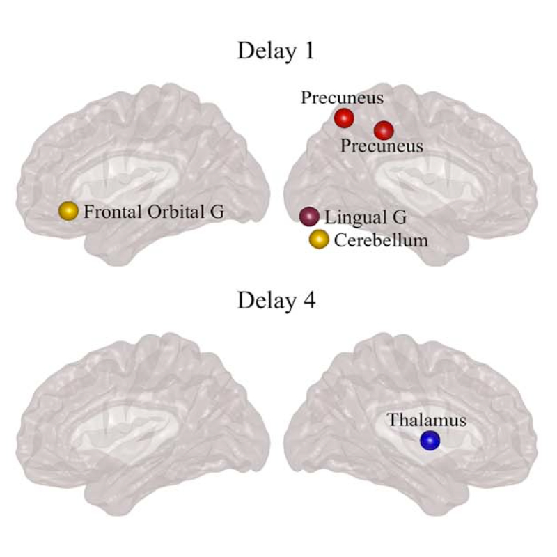
Differences between controls and PD participants in nodal network measures. (Image taken from the article.)Directed Brain Connectivity Identifies Widespread Functional Network Abnormalities in Parkinson’s Disease
Mite Mijalkov, Giovanni Volpe, Joana B Pereira
Cerebral Cortex, bhab237 (2021)
doi: 10.1093/cercor/bhab237
Parkinson’s disease (PD) is a neurodegenerative disorder characterized by topological abnormalities in large-scale functional brain networks, which are commonly analyzed using undirected correlations in the activation signals between brain regions. This approach assumes simultaneous activation of brain regions, despite previous evidence showing that brain activation entails causality, with signals being typically generated in one region and then propagated to other ones. To address this limitation, here, we developed a new method to assess whole-brain directed functional connectivity in participants with PD and healthy controls using antisymmetric delayed correlations, which capture better this underlying causality. Our results show that whole-brain directed connectivity, computed on functional magnetic resonance imaging data, identifies widespread differences in the functional networks of PD participants compared with controls, in contrast to undirected methods. These differences are characterized by increased global efficiency, clustering, and transitivity combined with lower modularity. Moreover, directed connectivity patterns in the precuneus, thalamus, and cerebellum were associated with motor, executive, and memory deficits in PD participants. Altogether, these findings suggest that directional brain connectivity is more sensitive to functional network differences occurring in PD compared with standard methods, opening new opportunities for brain connectivity analysis and development of new markers to track PD progression.