Vulnerability of locus coeruleus connections to aging and Alzheimer’s disease published in Alzheimer’s & Dementia

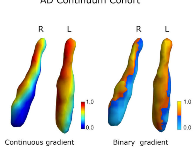
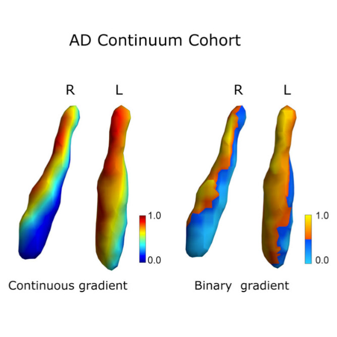
Anatomical gradients of the right (R) and left (L) LC in the AD continuum cohort (ADNI). (Image from the article.)
Vulnerability of locus coeruleus connections to aging and Alzheimer’s disease
Blanca Zufiria-Gerbolés, Daniel Vereb, Mite Mijalkov, Massimiliano Passaretti, Giovanni Volpe, Zhilei Xu, Thomas Hinault, Sara Garcia-Ptacek, Joana B. Pereira, for the Alzheimer’s Disease Neuroimaging Initiative
Alzheimer’s & Dementia 22, e71737 (2026)
DOI: 10.1002/alz.71737

INTRODUCTION
The locus coeruleus (LC) is important in coordinating communication between brain regions through its widespread connections. However, the organization of its connections, and its changes with aging and Alzheimer’s disease (AD), remains unclear.

METHODS
We mapped whole-brain white matter connections from the LC in two independent cohorts: one spanning the adult lifespan and another covering the AD continuum.

RESULTS
We identified a novel dorsal–ventral organization of LC connectivity that showed changes common to aging and AD or specific to AD, was linked to gene expression patterns, and was associated with cognitive performance and tau pathology in the entorhinal cortex. We also developed an imaging marker, LC gap, capturing LC connectivity deviations associated with slower tau accumulation in cognitively normal individuals at high risk for AD.

DISCUSSION
These findings provide new insights into LC connectivity in aging and AD, highlighting its potential role as a resilience marker against AD neurodegeneration.

Highlights

  • For the first time, we mapped the whole-brain white matter connections from the Locus Coeruleus (LC) to the rest of the brain across aging and Alzheimer’s disease (AD) continuum.
  • Revealed a novel dorsal–ventral organization LC connectivity that presents changes common to aging and AD or specific to AD.
  • LC connectivity patterns are associated with cognitive performance and tau pathology in the entorhinal cortex.
  • We introduced a novel marker capturing LC connectivity deviations and that is associated with tau accumulation in cognitively normal individuals at high risk for AD.
Mapping individual molecular connectomes in Alzheimer’s disease published in Alzheimer’s & Dementia

Diagnostic classification. (Image from the article.)
Mapping individual molecular connectomes in Alzheimer’s disease
Zhilei Xu, Mite Mijalkov, Jiawei Sun, Yu-Wei Chang, Arianna Sala, Giovanni Volpe, Mario Severino, Mattia Veronese, Sara Garcia-Ptacek, Joana B. Pereira, for the Alzheimer’s Disease Neuroimaging Initiative
Alzheimer’s & Dementia 22, e71310 (2026)
DOI: 10.1002/alz.71310

INTRODUCTION
Mapping individual differences is crucial to improve personalized medicine approaches in Alzheimer’s disease (AD), which is characterized by strong inter-individual variability in the accumulation patterns of tau and amyloid beta pathology.

METHODS
We assess the progression of AD across the disease continuum by building individual molecular connectomes using longitudinal positron emission tomography (PET) data.

RESULTS
We demonstrate that these connectomes constitute a unique fingerprint, capable of identifying a single individual from a large group of subjects. Alterations in the connectomes discriminate different diagnostic groups and predict cognitive decline to a higher extent than conventional PET measures. We introduce a novel gene-specific transcription network analysis that linked individual tau and amyloid connectomes to a common transcriptomic profile of apoptosis, with the tau connectome being specifically related to pyrimidine metabolism, and the amyloid connectome to histone acetylation.

DISCUSSION
Individual molecular connectome mapping provides a novel and sensitive framework to monitor AD progression.

Highlights

  • Individual molecular connectomes constitute a unique fingerprint, capable of identifying a single individual from a large group of subjects.
  • Alterations in individual molecular connectomes significantly increase both across the Alzheimer’s disease (AD) continuum and over time.
  • Alterations in individual molecular connectomes discriminate different diagnostic groups and predict cognitive decline to a higher extent than conventional positron emission tomography measures.
  • Susceptibilities of individual tau and amyloid connectomes to AD are linked to a common transcriptomic profile of apoptosis, with the tau connectome being specifically related to pyrimidine metabolism, and the amyloid connectome to histone acetylation.

Tracking early cognitive decline in preclinical AD with brain MRI similarity published in Alzheimer’s & Dementia

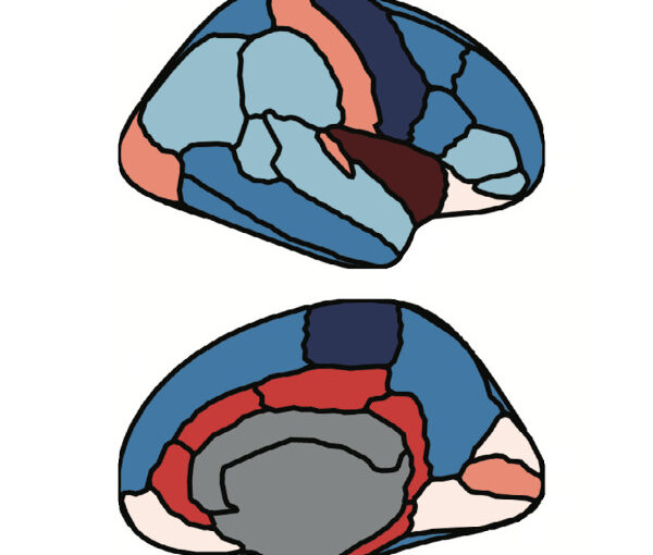
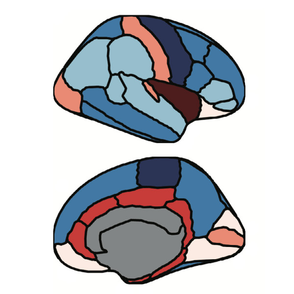
Parcellation of the brain cortex. (Image from the article.)
Tracking early cognitive decline in preclinical AD with brain MRI similarity
Jiawei Sun, Blanca Zufiria-Gerbolés, Massimiliano Passaretti, Giovanni Volpe, Mite Mijalkov, Joana B. Pereira, for the Alzheimer’s Disease Neuroimaging Initiative
Alzheimer’s & Dementia 22, e71170 (2026)
DOI: 10.1002/alz.71170

INTRODUCTION
Early detection of neuroanatomical changes in preclinical Alzheimer’s disease (AD) is critical for timely intervention. However, conventional magnetic resonance imaging (MRI) and fluid biomarkers often lack sensitivity to subtle structural alterations in early disease stages.

METHODS
To identify early brain alterations, we applied a perturbation-based brain similarity approach to cognitively normal participants from Alzheimer’s Disease Neuroimaging Initiative (ADNI) and Open Access Series of Imaging Studies (OASIS), stratified by amyloid status. We evaluated its predictive performance for cognition and diagnostic conversion against cortical thickness, volumetric MRI, and fluid biomarkers.

RESULTS
In both cohorts, brain similarity consistently outperformed other biomarkers across cognitive domains and amyloid groups. It also achieved superior accuracy in predicting clinical conversion and exhibited associations with cytoarchitectural organization.

DISCUSSION
These findings highlight brain similarity as a sensitive marker of early neuroanatomical disruption in AD. Its ability to detect subtle structural changes before overt atrophy underscores its potential for early disease monitoring and treatment assessment in preclinical AD trials.

Highlights

  • Brain similarity captures early brain changes in preclinical Alzheimer’s disease (AD).
  • Brain similarity outperforms conventional biomarkers such as cortical thickness, volume measures, and fluid biomarkers in predicting cognitive decline.
  • Brain similarity predicts conversion to mild cognitive impairment and AD more accurately than traditional imaging markers, and its predictive performance is further improved when combined with fluid biomarkers.
  • Brain similarity captures structural disruptions associated with cortical layer II of the cytoarchitectonic lamina of human neocortex.