Martin Selin, Antonio Ciarlo, Giuseppe Pesce, Lars Bengtsson, Joan Camunas-Soler, Vinoth Sundar Rajan, Fredrik Westerlund, L. Marcus Wilhelmsson, Isabel Pastor, Felix Ritort, Steven B. Smith, Carlos Bustamante, and Giovanni Volpe
Date: 11th March 2026
Time: 18:00 – 20:00
Place: Aula Medica, Karolinska Institute, Solna
Conference Protein Folding in Real Time, 11-13 March 2026, Stockholm, Sweden
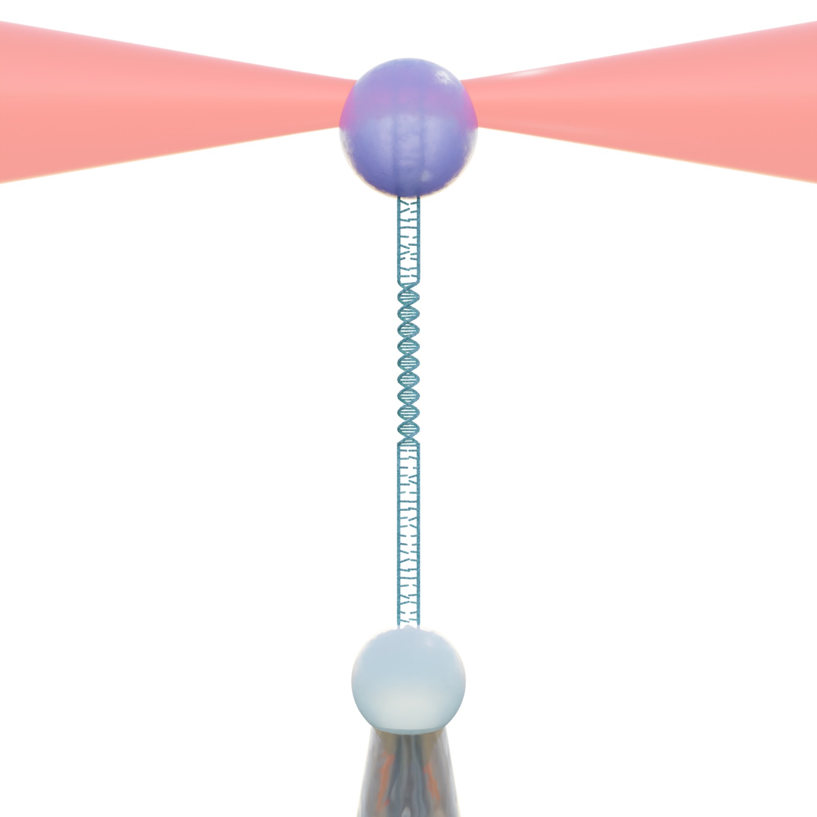
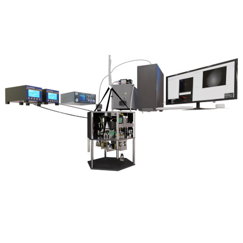
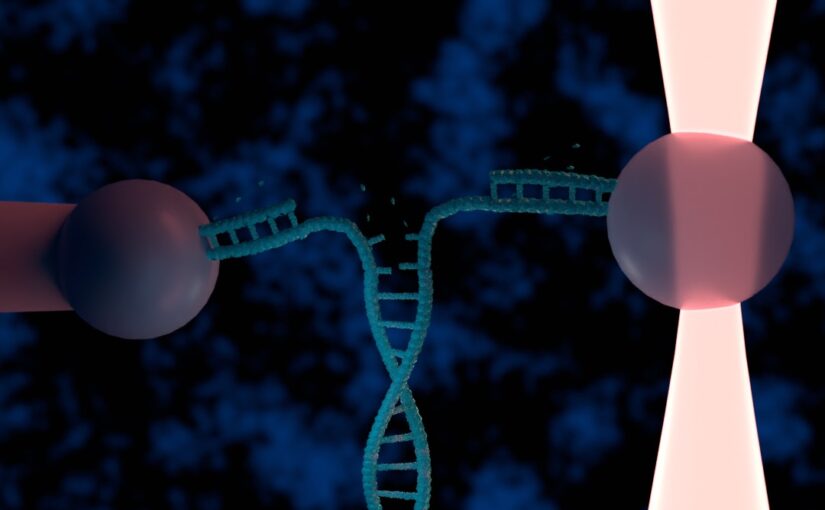
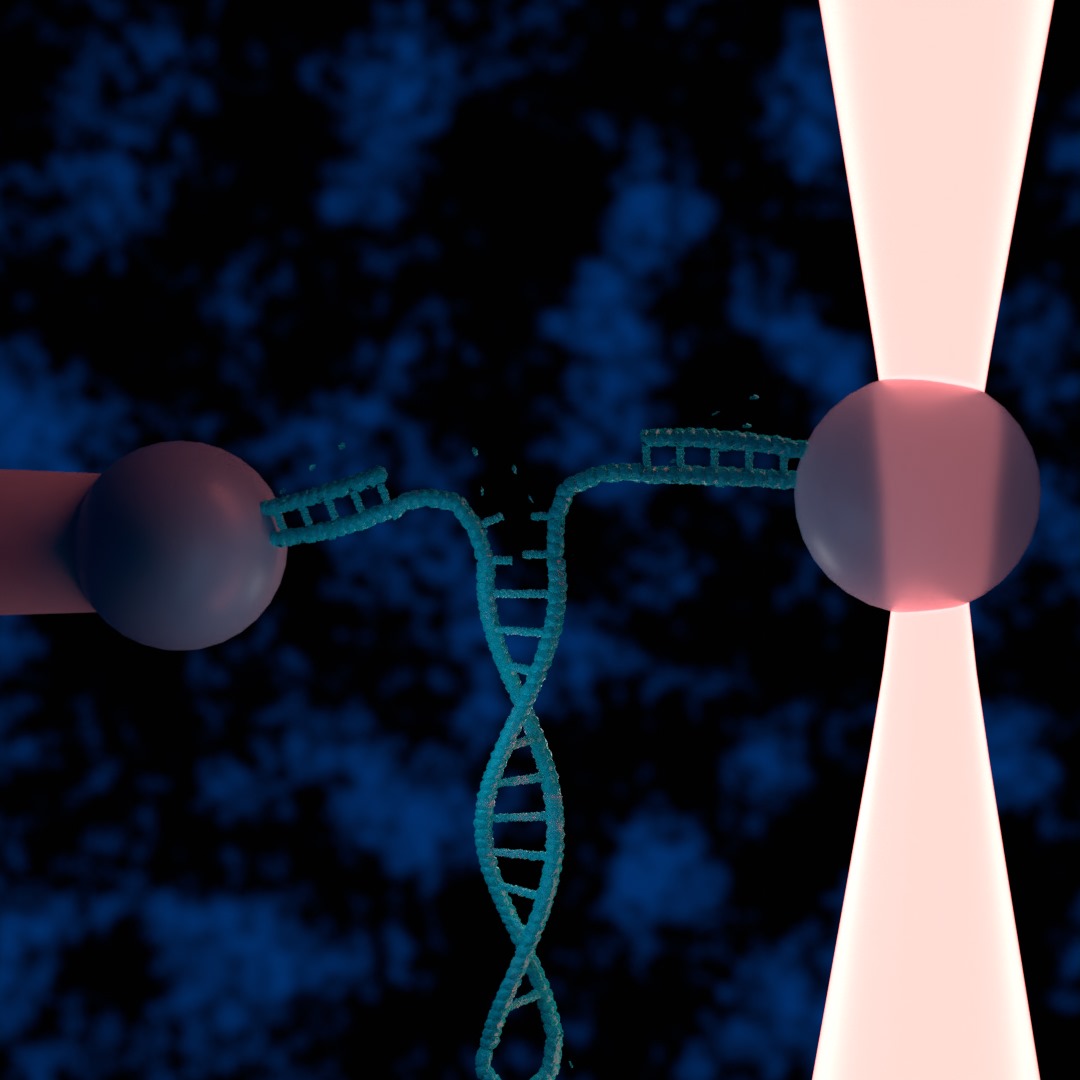
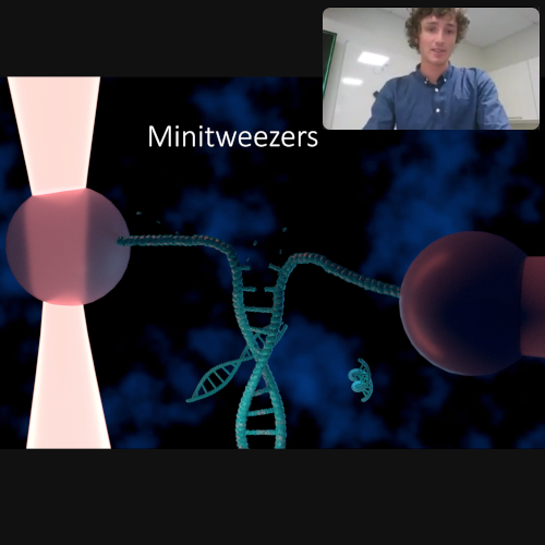
Single-molecule studies are vital for understanding fundamental biological processes, including protein folding, DNA transcription, and replication. However, performing these experiments manually on individual molecules is notoriously time-consuming and costly. To address this challenge, we have developed a fully autonomous single-molecule force spectroscopy platform by integrating a custom-built optical tweezers instrument with real-time deep-learning-based image analysis and adaptive control protocols. Our system achieves human-level throughput in terms of experiments per hour while remaining robust enough to operate continuously for hours without intervention. We demonstrate the versatility of our platform by having it perform DNA pulling experiments fully autonomously. By making the software open source we democratize high-throughput data collection in single-molecule biophysics, paving the way for merging single-molecule studies with large-scale, data-driven approaches—ultimately enabling new insights into the dynamic, transient states of complex biological systems.