Presentation by G. V. P. Kumar, 24 November 2021

Thermoplasmonic Tweezers: Probing single-molecules and more
G. V. Pavan Kumar
IISER, Pune, India.
24 November 2021
Online

In this presentation, we will discuss two specific issues: How to perform single-molecule surface enhanced Raman scattering (SERS) in an optothermal trap? and how to design optothermal fields to trap and interrogate molecules and colloids in a fluid?

In recent years, performing SERS in optical traps has emerged as an important development in nano- and bio-photonics. To this end, tweezer techniques based on surface-plasmons facilitate deeper optical potentials at sub-wavelength scales, and simultaneously provide enhanced electric and optothermal fields. In this
presentation, we will discuss various strategies developed in my laboratory to perform single-molecule SERS in optical and plasmonic tweezer platforms. Specifically, we will highlight some thermoplasmonic effects and directionality aspects of the tweezer platforms in metallic thin film and some plasmonic nano-architectures.

Short bio:

G.V. Pavan Kumar is an associate professor of physics at the Indian Institute of Science Education and Research (IISER), Pune, India.
He obtained his PhD from JNCASR, Bangalore. Subsequently he was a postdoctoral fellow at ICFO-Barcelona and Purdue University, before joining IISER in 2010.
His current research interests are optical, optothermal and nanophotonic forces and their utility in probing single molecules and soft-matter systems at micro and nanoscale.
To this end, his lab has been interfacing optical tweezer platforms with a variety of optical spectroscopy and microscopy tools.
He blogs on topics related to science: https://backscattering.wordpress.com/
Invited Presentation by G. Volpe at FiO LS, 4 November 2021

DeepTrack 2.0 Logo. (Image from DeepTrack 2.1 Project)
DeepTrack 2.1: A Framework for Deep Learning for Microscopy
Giovanni Volpe
Invited Presentation at Frontiers in Optics + Laser Science
Online
4 November 2021
4:00 PM

We present DeepTrack 2.0, a software to design, train, and validate deep-learning solutions for digital microscopy. We demonstrate it for applications from particle localization, tracking, and characterization, to cell counting and classification, to virtual staining.

Link: FTh6A.3

Seminar by D. Midtvedt at Freie Universität Berlin, 29 October 2021

DeepTrack 2.0 Logo. (Image from DeepTrack 2.0 Project)
Quantitative digital microscopy enhanced by deep learning
Daniel Midtvedt
(online at) Freie Universität Berlin, Germany
29 October 2021

Video microscopy has a long history of providing insight and breakthroughs for a broad range of disciplines, from physics to biology. Image analysis to extract quantitative information from video microscopy data has traditionally relied on algorithmic approaches, which are often difficult to implement, time-consuming, and computationally expensive. Recently, alternative data-driven approaches using deep learning have greatly improved quantitative digital microscopy, potentially offering automatized, accurate, and fast image analysis.
However, the combination of deep learning and video microscopy remains underutilized primarily due to the steep learning curve involved in developing custom deep-learning solutions. To overcome this issue, we recently introduced a software, DeepTrack 2.0, to design, train, and validate deep-learning solutions for digital microscopy.
In this talk, I will show how this software can be used in a broad range of applications, from particle localization, tracking, and characterization, to cell counting and classification. Thanks to its user-friendly graphical interface, DeepTrack 2.0 can be easily customized for user-specific applications, and thanks to its open-source, object-oriented programing, it can be easily expanded to add features and functionalities, potentially introducing deep-learning-enhanced video microscopy to a far wider audience.

Invited Talk by G. Volpe at Microscopies and Spectroscopies: Accessing the Nanoscale, 28 October 2021

DeepTrack 2.0 Logo. (Image from DeepTrack 2.0 Project)
Quantitative Digital Microscopy with Deep Learning
Giovanni Volpe
Invited Talk at the XXXVI Trobades Cientifíques de la Mediterránia – Josep Miquel Vidal
Microscopies and Spectroscopies: Accessing the Nanoscale
Menorca, Spain
28 October 2021
11:40 AM

Video microscopy has a long history of providing insights and breakthroughs for a broad range of disciplines, from physics to biology. Image analysis to extract quantitative information from video microscopy data has traditionally relied on algorithmic approaches, which are often difficult to implement, time consuming, and computationally expensive. Recently, alternative data-driven approaches using deep learning have greatly improved quantitative digital microscopy, potentially offering automatized, accurate, and fast image analysis. However, the combination of deep learning and video microscopy remains underutilized primarily due to the steep learning curve involved in developing custom deep-learning solutions. To overcome this issue, we introduce a software, DeepTrack 2.0, to design, train and validate deep- learning solutions for digital microscopy. We use it to exemplify how deep learning can be employed for a broad range of applications, from particle localization, tracking and characterization to cell counting and classification. Thanks to its user- friendly graphical interface, DeepTrack 2.0 can be easily customized for user-specific applications, and, thanks to its open-source object-oriented programming, it can be easily expanded to add features and functionalities, potentially introducing deep-learning-enhanced video microscopy to a far wider audience.

Presentation by S. Olsson, 20 October 2021

Machine Learning for Molecular dynamics — Why bother?
Simon Olsson
Chalmers University of Technology
20 October 2021
Online

With faster compute-infrastructures, molecular simulations play an increasingly important role in the basic sciences and application areas such as drug and materials design. Simultaneously, machine learning and artificial intelligence are receiving increased attention due to increasing volumes of data generated both inside and outside of science. In this talk, I will talk about a few applications of these technologies in molecular simulation, focusing on biomolecular simulations [1,2]

[1] Olsson & Noé ”Dynamic Graphical Models of Molecular Kinetics” Proc. Natl. Acad. Sci. U.S.A. (2019) doi: 10.1073/pnas.1901692116.
[2] Noe†, Olsson, Köhler, Wu ”Boltzmann Generators: Sampling Equilibrium States of Many-Body Systems with Deep Learning” Science (2019). 365, eaaw1147. doi:10.1126/science.aaw1147.

Link: http://www.cse.chalmers.se/~simonols/

Keynote Talk by G. Volpe at CIIBBI, 15 October 2021

DeepTrack 2.0 Logo. (Image from DeepTrack 2.0 Project)
Deep Learning for Microscopy with Biomedical Applications
Giovanni Volpe
Keynote Talk at the 2nd International Congress of Biomedical Engineering and Bioengineering
Online
15 October 2021
14:00 CEST

Video microscopy has a long history of providing insights and breakthroughs for a broad range of disciplines, from physics to biology. Image analysis to extract quantitative information from video microscopy data has traditionally relied on algorithmic approaches, which are often difficult to implement, time consuming, and computationally expensive. Recently, alternative data-driven approaches using deep learning have greatly improved quantitative digital microscopy, potentially offering automatized, accurate, and fast image analysis. However, the combination of deep learning and video microscopy remains underutilized primarily due to the steep learning curve involved in developing custom deep-learning solutions. To overcome this issue, we introduce a software, DeepTrack 2.0, to design, train and validate deep-learning solutions for digital microscopy. We use it to exemplify how deep learning can be employed for a broad range of applications, from particle localization, tracking and characterization to cell counting and classification. Thanks to its user-friendly graphical interface, DeepTrack 2.0 can be easily customized for user-specific applications, and, thanks to its open-source object-oriented programming, it can be easily expanded to add features and functionalities, potentially introducing deep-learning-enhanced video microscopy to a far wider audience.

Invited Talk by G. Volpe at 106 RAFA, 15 October 2021

Deep learning for microscopy, optical trapping, and active matter
Giovanni Volpe
Invited Talk for 106 RAFA – Reunión de la Asociación de Física de Argentina – División Materia Blanda
15 October 2021
12:00 PM

After a brief overview of artificial intelligence, machine learning and deep learning, I will present a series of recent works in which we have employed deep learning for applications in photonics and active matter. In particular, I will explain how we employed deep learning to enhance digital video microscopy, to estimate the properties of anomalous diffusion, to characterize microscopic force fields, to improve the calculation of optical forces, and to characterize nanoparticles. Finally, I will provide an outlook for the application of deep learning in photonics and active matter.

Invited Talk by G. Volpe at Venice meeting on Fluctuations in small complex systems V, 7 October 2021

RANDI architecture to classify the model underlying anomalous diffusion.
Measuring Anomalous Diffusion with Deep Learning
Giovanni Volpe
Invited Talk at Venice meeting on Fluctuations in small complex systems V
Palazzo Franchetti, Venezia, Italy
7 October 2021, 16:30 CEST

Countless systems in biology, physics, and finance undergo diffusive dynamics. Many of these systems, including biomolecules inside cells, active matter systems and foraging animals, exhibit anomalous dynamics where the growth of the mean squared displacement with time follows a power law with an exponent that deviates from 1. When studying time series recording the evolution of these systems, it is crucial to precisely measure the anomalous exponent and confidently identify the mechanisms responsible for anomalous diffusion. These tasks can be overwhelmingly difficult when only few short trajectories are available, a situation that is common in the study of non-equilibrium and living systems. Here, we present a data-driven method to analyze single anomalous diffusion trajectories employing recurrent neural networks, which we name RANDI. We show that our method can successfully infer the anomalous exponent, identify the type of anomalous diffusion process, and segment the trajectories of systems switching between different behaviors. We benchmark our performance against the state-of-the art techniques for the study of single short trajectories that participated in the Anomalous Diffusion (AnDi) challenge. Our method proved to be the most versatile method, being the only one to consistently rank in the top 3 for all tasks proposed in the AnDi challenge.

Invited Talk by G. Volpe at Machine Learning and Automated Experiment in Scanning Probe Microscopy, 4 October 2021

DeepTrack 2.0 Logo. (Image from DeepTrack 2.0 Project)
Quantitative Digital Microscopy with Deep Learning
Giovanni Volpe
Invited Talk at the Virtual school “Machine Learning and Automated Experiment in Scanning Probe Microscopy”
Online
October 4-7, 2021
11:20 AM

Video microscopy has a long history of providing insights and breakthroughs for a broad range of disciplines, from physics to biology. Image analysis to extract quantitative information from video microscopy data has traditionally relied on algorithmic approaches, which are often difficult to implement, time consuming, and computationally expensive. Recently, alternative data-driven approaches using deep learning have greatly improved quantitative digital microscopy, potentially offering automatized, accurate, and fast image analysis. However, the combination of deep learning and video microscopy remains underutilized primarily due to the steep learning curve involved in developing custom deep-learning solutions. To overcome this issue, we introduce a software, DeepTrack 2.0, to design, train and validate deep- learning solutions for digital microscopy. We use it to exemplify how deep learning can be employed for a broad range of applications, from particle localization, tracking and characterization to cell counting and classification. Thanks to its user- friendly graphical interface, DeepTrack 2.0 can be easily customized for user-specific applications, and, thanks to its open-source object-oriented programming, it can be easily expanded to add features and functionalities, potentially introducing deep-learning-enhanced video microscopy to a far wider audience.

Invited Talk by G. Volpe at ICTP, 8 September 2021

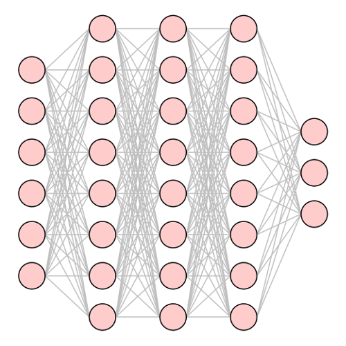
Neural net with input layer (left), dense internal layers, and output layer (right). (Image from the article Machine Learning for Active Matter)
Machine Learning for Active Matter: Opportunities and Challenges

Giovanni Volpe
Invited Talk
(online at) ICTP, Trieste, Italy
8 September 2021, 11:30 CEST

Machine-learning methods are starting to shape active-matter research. Which new trends will this start? Which new groundbreaking insight and applications can we expect? More fundamentally, what can this contribute to our understanding of active matter? Can this help us to identify unifying principles and systematise active matter? This presentation addresses some of these questions with some concrete examples, exploring how machine learning is steering active matter towards new directions, offering unprecedented opportunities and posing practical and fundamental challenges. I will illustrate some most successful recent applications of machine learning to active matter with a slight bias towards work done in my research group: enhancing data acquisition and analysis; providing new data-driven models; improving navigation and search strategies; offering insight into the emergent dynamics of active matter in crowded and complex environments. I will discuss the opportunities and challenges that are emerging: implementing feedback control; uncovering underlying principles to systematise active matter; understanding the behaviour, organisation and evolution of biological active matter; realising active matter with embodied intelligence. Finally, I will highlight how active matter and machine learning can work together for mutual benefit.

Links:
Conference: Statistical Physics of Complex Systems | (smr 3624)
Program of Day 1 (8 September)