Tracking early cognitive decline in preclinical AD with brain MRI similarity published in Alzheimer’s & Dementia

Parcellation of the brain cortex. (Image from the article.)
Tracking early cognitive decline in preclinical AD with brain MRI similarity
Jiawei Sun, Blanca Zufiria-Gerbolés, Massimiliano Passaretti, Giovanni Volpe, Mite Mijalkov, Joana B. Pereira, for the Alzheimer’s Disease Neuroimaging Initiative
Alzheimer’s & Dementia 22, e71170 (2026)
DOI: 10.1002/alz.71170

INTRODUCTION
Early detection of neuroanatomical changes in preclinical Alzheimer’s disease (AD) is critical for timely intervention. However, conventional magnetic resonance imaging (MRI) and fluid biomarkers often lack sensitivity to subtle structural alterations in early disease stages.

METHODS
To identify early brain alterations, we applied a perturbation-based brain similarity approach to cognitively normal participants from Alzheimer’s Disease Neuroimaging Initiative (ADNI) and Open Access Series of Imaging Studies (OASIS), stratified by amyloid status. We evaluated its predictive performance for cognition and diagnostic conversion against cortical thickness, volumetric MRI, and fluid biomarkers.

RESULTS
In both cohorts, brain similarity consistently outperformed other biomarkers across cognitive domains and amyloid groups. It also achieved superior accuracy in predicting clinical conversion and exhibited associations with cytoarchitectural organization.

DISCUSSION
These findings highlight brain similarity as a sensitive marker of early neuroanatomical disruption in AD. Its ability to detect subtle structural changes before overt atrophy underscores its potential for early disease monitoring and treatment assessment in preclinical AD trials.

Highlights

  • Brain similarity captures early brain changes in preclinical Alzheimer’s disease (AD).
  • Brain similarity outperforms conventional biomarkers such as cortical thickness, volume measures, and fluid biomarkers in predicting cognitive decline.
  • Brain similarity predicts conversion to mild cognitive impairment and AD more accurately than traditional imaging markers, and its predictive performance is further improved when combined with fluid biomarkers.
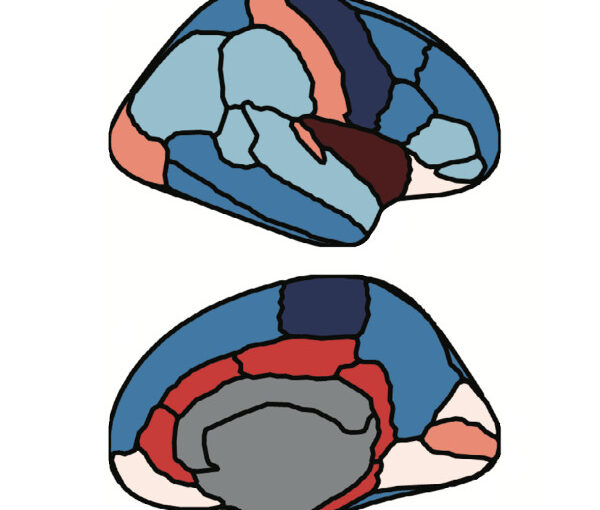
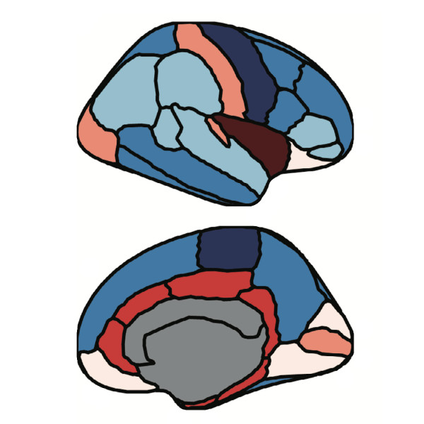
  • Brain similarity captures structural disruptions associated with cortical layer II of the cytoarchitectonic lamina of human neocortex.

Workshop by Y.-W. Chang at NEMES 2025, Gothenburg, 26 September 2025

Massimiliano Passaretti (left) and Yu-Wei Chang (right) at NEME 2025. (Photo courtesy of Clarion Hotel Draken.)
Graph theory and deep learning pipelines
Yu-Wei Chang, Massimiliano Passaretti
NEMES 2025, 24-26 September, 2025
Date: 25 September 2025
Time: 12:45 – 14:00
Place: Clarion Hotel Draken

This workshop begins with a practical introduction to graph theory, then guides participants through BRAPH 2 to build connectomes, compute graph measures, and run group comparisons, followed by a hands-on deep-learning pipeline. It demonstrates a unified GUI/command-line workflow, a unique architecture of BRAPH 2, helping participants move smoothly from the GUI to scripts. This workshop also guides participants to reproduce multiplex and deep-learning results on their computers from the BRAPH 2 bioRxiv preprint.
 

Computational memory capacity predicts aging and cognitive decline published in Nature Communications

Memory capacity in aging. A Brain reservoir computing architecture with uniform random signals applied to all nodes. (Image from the article.)
Computational memory capacity predicts aging and cognitive decline
Mite Mijalkov, Ludvig Storm, Blanca Zufiria-Gerbolés, Dániel Veréb, Zhilei Xu, Anna Canal-Garcia, Jiawei Sun, Yu-Wei Chang, Hang Zhao, Emiliano Gómez-Ruiz, Massimiliano Passaretti, Sara Garcia-Ptacek, Miia Kivipelto, Per Svenningsson, Henrik Zetterberg, Heidi Jacobs, Kathy Lüdge, Daniel Brunner, Bernhard Mehlig, Giovanni Volpe, Joana B. Pereira
Nature Communications 16, 2748 (2025)
doi: 10.1038/s41467-025-57995-0

Memory is a crucial cognitive function that deteriorates with age. However, this ability is normally assessed using cognitive tests instead of the architecture of brain networks. Here, we use reservoir computing, a recurrent neural network computing paradigm, to assess the linear memory capacities of neural-network reservoirs extracted from brain anatomical connectivity data in a lifespan cohort of 636 individuals. The computational memory capacity emerges as a robust marker of aging, being associated with resting-state functional activity, white matter integrity, locus coeruleus signal intensity, and cognitive performance. We replicate our findings in an independent cohort of 154 young and 72 old individuals. By linking the computational memory capacity of the brain network with cognition, brain function and integrity, our findings open new pathways to employ reservoir computing to investigate aging and age-related disorders.