Sophia Simon is a bachelor student at the Freie Universität of Berlin. She will do her summer internship at the Soft Matter Lab from July 21 to September 27, 2019, with a grant from DAAD (Deutscher Akademischer Austauschdienst). She will work on the tunability of critical Casimir forces in critical mixtures.
News
Anomalous Diffusion Measurement with Neural Networks published in Phys Rev E
Measurement of Anomalous Diffusion Using Recurrent Neural Networks
Stefano Bo, Falko Schmidt, Ralf Eichborn & Giovanni Volpe
Physical Review E 100(1), 010102(R) (2019)
doi: 10.1103/PhysRevE.100.010102
arXiv: 1905.02038
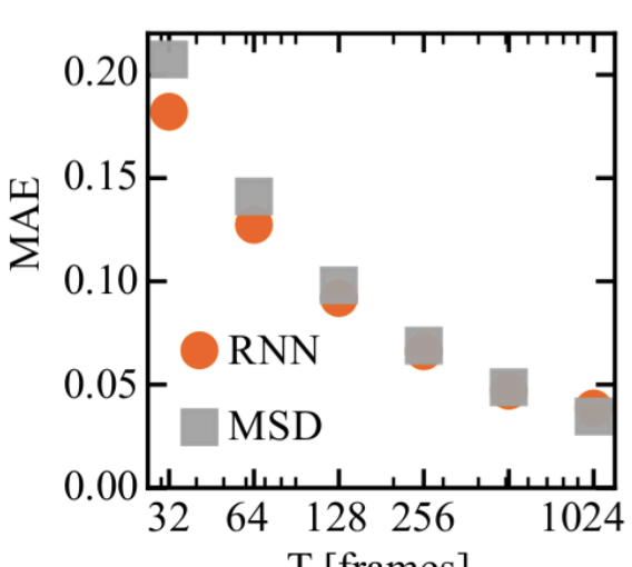
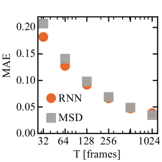
Anomalous diffusion occurs in many physical and biological phenomena, when the growth of the mean squared displacement (MSD) with time has an exponent different from one. We show that recurrent neural networks (RNN) can efficiently characterize anomalous diffusion by determining the exponent from a single short trajectory, outperforming the standard estimation based on the MSD when the available data points are limited, as is often the case in experiments. Furthermore, the RNN can handle more complex tasks where there are no standard approaches, such as determining the anomalous diffusion exponent from a trajectory sampled at irregular times, and estimating the switching time and anomalous diffusion exponents of an intermittent system that switches between different kinds of anomalous diffusion. We validate our method on experimental data obtained from sub-diffusive colloids trapped in speckle light fields and super-diffusive microswimmers.
Influence of Sensorial Delay on Clustering and Swarming published in Phys. Rev. E
Influence of Sensorial Delay on Clustering and Swarming
Rafal Piwowarczyk, Martin Selin, Thomas Ihle & Giovanni Volpe
Physical Review E 100(1), 012607 (2019)
doi: 10.1103/PhysRevE.100.012607
arXiv: 1803.06026
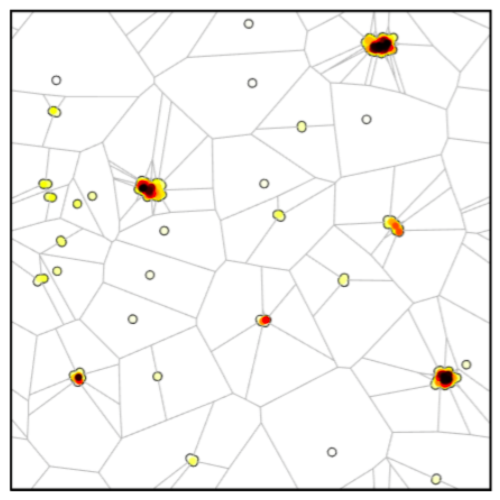
We show that sensorial delay alters the collective motion of self-propelling agents with aligning interactions: In a two-dimensional Vicsek model, short delays enhance the emergence of clusters and swarms, while long or negative delays prevent their formation. In order to quantify this phenomenon, we introduce a global clustering parameter based on the Voronoi tessellation, which permits us to efficiently measure the formation of clusters. Thanks to its simplicity, sensorial delay might already play a role in the organization of living organisms and can provide a powerful tool to engineer and dynamically tune the behavior of large ensembles of autonomous robots.
Falko Schmidt attends the 69th Lindau Nobel laureate meeting
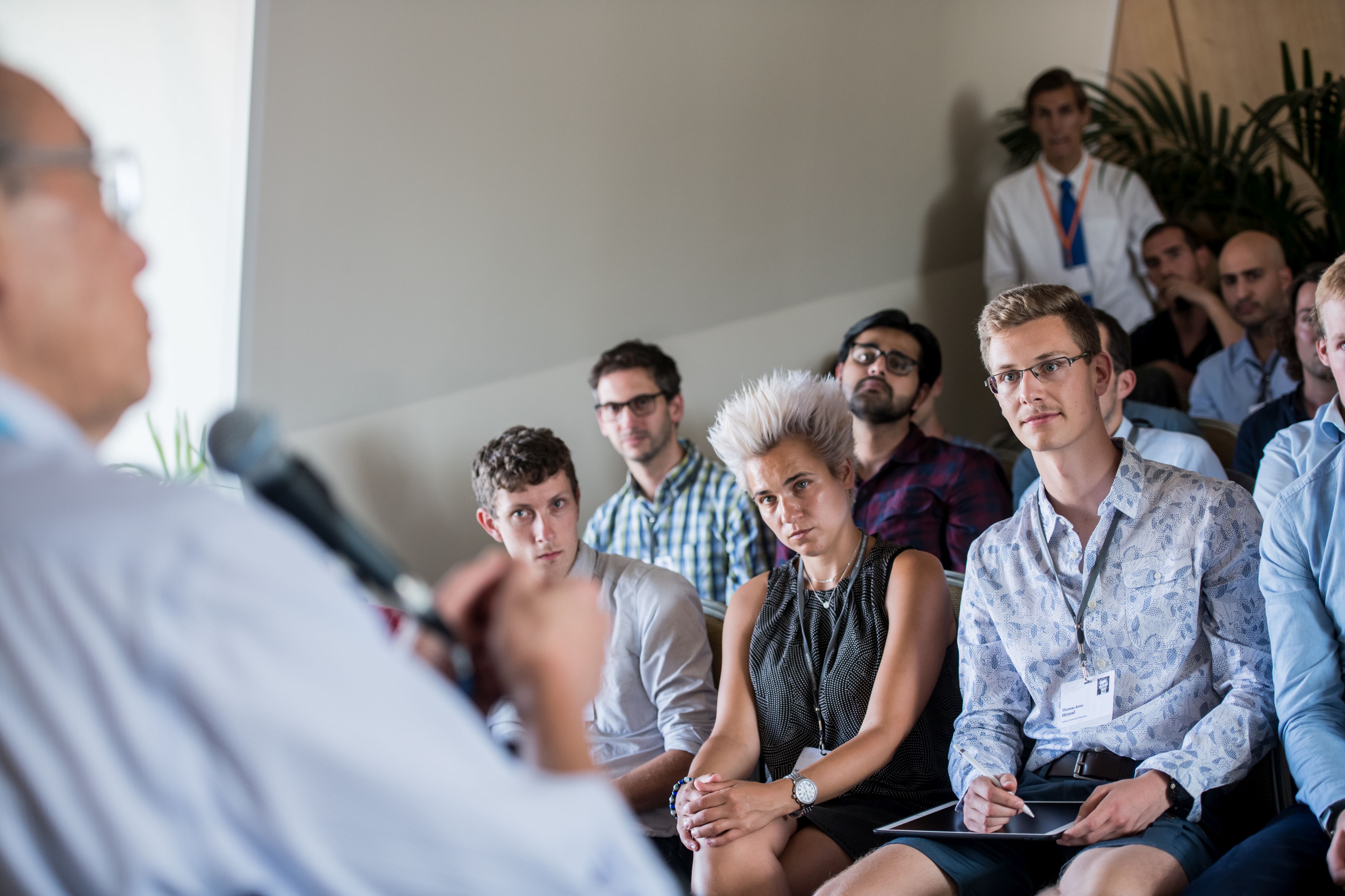
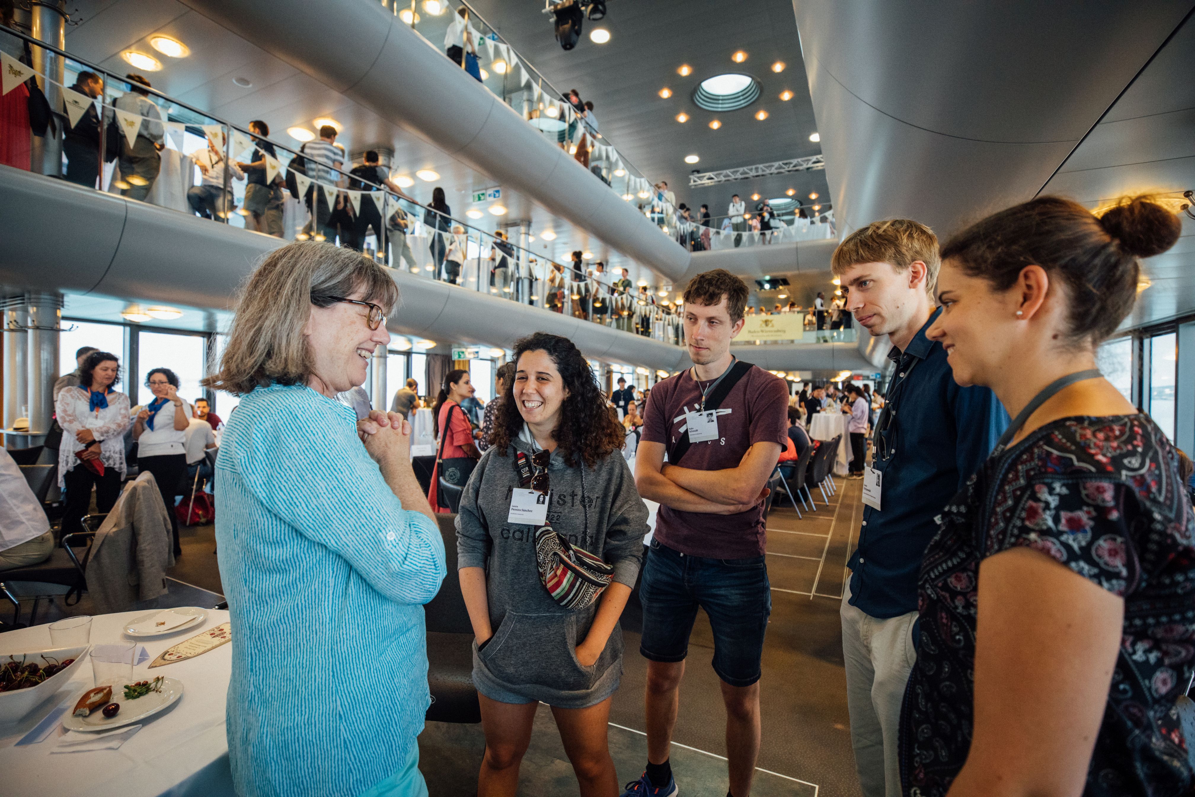
Picture/Credit: Julia Nimke/Lindau Nobel Laureate Meetings
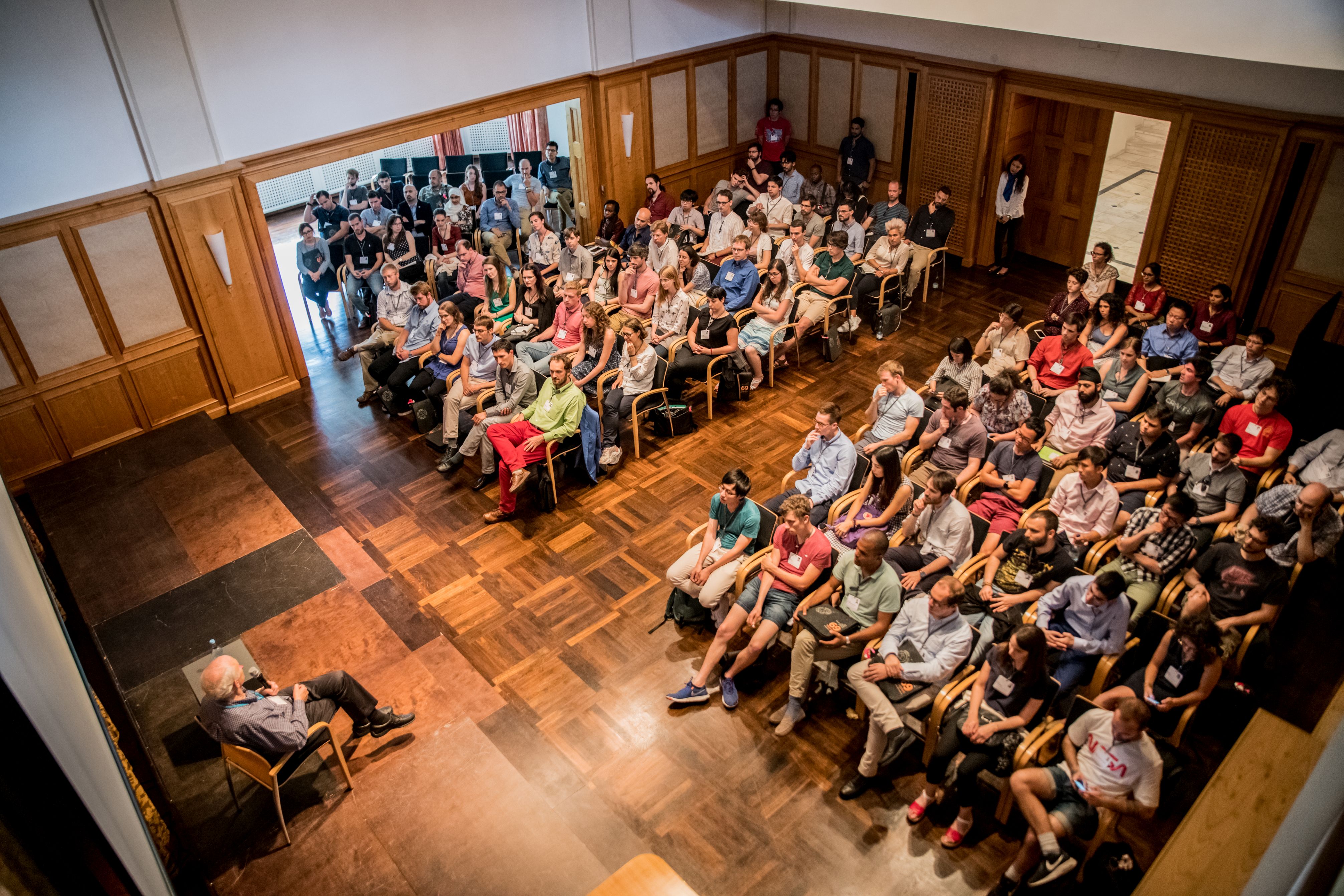
Falko Schmidt, and Jalpa Soni have been selected to attain the 69th Lindau Nobel Laureate meeting in Lindau, Germany from the 30th June till 5th July 2019.
The Lindau meeting is a platform where 600 young scientists around the world meet former Nobel laureates (as well as Turing-award winners). There they can exchange scientific ideas and experiences, inspire each other and connect for a more interdisciplinary scientific community. These are the three incentives that make this meeting a unique experience.
Falko Schmidt had the privilege to attend it and shares the following insight:
“For me, the Lindau meeting was a unique experience where I was able to meet peers across many disciplines, share ideas and experiences beyond my field of active matter and received much feedback on career choices and daily life as a PhD. Especially fruitful were the many possibilities to engage with senior scientists such as the Nobel laureates which with their humour, insight and advice deepened my passion about science. Personally, I would consider my best encounters with Steven Chu and William Phillips (Nobel Prize in Physics in 1997 on laser cooling), Donna Strickland (Nobel Prize in Physics in 2018 on ultra-fast lasers), and Stefan Hell (Nobel Prize in Chemistry in 2014 on super-resolution microscopy). I am very grateful for the possibility of attending this meeting and would like to thank the Lindau Nobel committee and Söderbergs Foundation who were selecting and sponsoring me.
From now on, in times of struggle, I will always look back to this meeting and remember why we all love doing science.”
Invited talk by G. Volpe at MARSS19, Espoo, 2 July 2019
Deep Learning for Measurement and Manipulation on the Microscale
Giovanni Volpe
Invited talk in the Special session on “Similarities and differences between biological and artificial micro robots” at MARSS2019
Espoo, Finland
1-5 July 2019
Intracavity Optical Trapping published in Nature Commun.
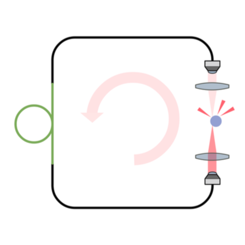
Intracavity optical trapping of microscopic particles in a ring-cavity fiber laser
Fatemeh Kalantarifard, Parviz Elahi, Ghaith Makey, Onofrio M. Maragò, F. Ömer Ilday & Giovanni Volpe
Nature Communications 10, 2683 (2019)
doi: 10.1038/s41467-019-10662-7
arXiv: 1808.07831
Standard optical tweezers rely on optical forces arising when a focused laser beam interacts with a microscopic particle: scattering forces, pushing the particle along the beam direction, and gradient forces, attracting it towards the high-intensity focal spot. Importantly, the incoming laser beam is not affected by the particle position because the particle is outside the laser cavity. Here, we demonstrate that intracavity nonlinear feedback forces emerge when the particle is placed inside the optical cavity, resulting in orders-of-magnitude higher confinement along the three axes per unit laser intensity on the sample. This scheme allows trapping at very low numerical apertures and reduces the laser intensity to which the particle is exposed by two orders of magnitude compared to a standard 3D optical tweezers. These results are highly relevant for many applications requiring manipulation of samples that are subject to photodamage, such as in biophysics and nanosciences.
Fatemeh Kalantarifard defended her PhD Thesis on 10 June 2019. Congrats!
Fatemeh Kalantarifard defended her PhD Thesis on 10 June 2019 in the Department of Physics Seminar Room SA-240 – Bilkent University.
Her Ph.D. Thesis Defense was live streamed on 10 June 2019 at 15:30 CEST in the Raven & Fox room.
Assoc. Prof. Ömer Ilday (UNAM, Bilkent University), Assoc. Prof. Alpan Bek (Middle-East Technical University), Assist. Prof. Burcin Ünlü (Bogazici University), Dr. Seymour Jahangirov (UNAM), Prof. Oguz Gülseren (Bilkent University) and Assist. Prof. Giovanni Volpe (Bilkent University) will be the thesis committee members.
Thesis title: Intra-cavity optical trapping with fiber laser
Thesis abstract: Standard optical tweezers rely on optical forces arising when a focused laser beam interacts with a microscopic particle: scattering forces, pushing the particle along the beam direction, and gradient forces, attracting it towards the high-intensity focal spot. Importantly, the incoming laser beam is not affected by the particle position because the particle is outside the laser cavity. Here, we demonstrate that intra-cavity nonlinear feedback forces emerge when the particle is placed inside the optical cavity, resulting in orders-of-magnitude higher confinement along the three axes per unit laser intensity on the sample. This scheme allows trapping at very low numerical apertures and reduces the laser intensity to which the particle is exposed by two orders of magnitude compared to a standard 3D optical tweezers. These results are highly relevant for many applications requiring manipulation of samples that are subject to photodamage, such as in biophysics and nano-sciences.
Thesis Advisor Giovanni Volpe, Department of Physics, Bilkent University
Place: Physics Department seminar room (SA240), Bilkent University
Time: 10 June, 2019, 16:30 TRT (Turkey Time)
LIVE STREAMING:
Place: Meeting room Raven & Fox, Gothenburg University
Time: 10 June, 2019, 15:30 CEST
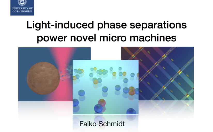
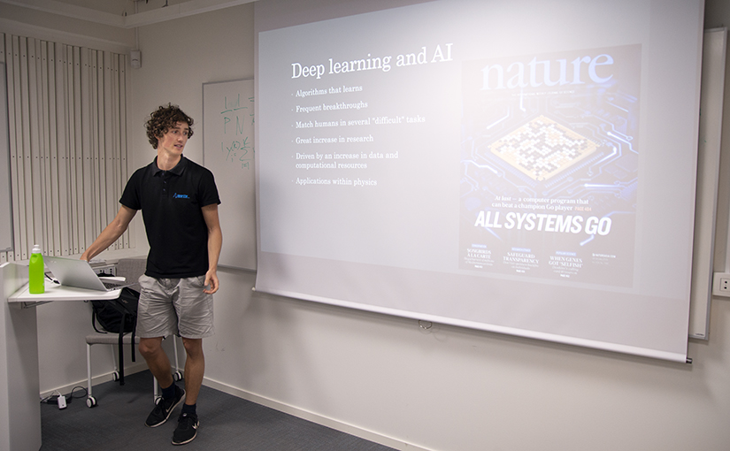
Falko Schmidt presented his PhD half-time seminar
About mid-way through his PhD, Falko Schmidt presented his past research activities and gave an outlook on his future projects. The topics range from miniaturised machines to self-assembled active molecules activated by light to machine-learning techniques to better characterise dynamical behaviour of microscopic systems.
The seminar will be held at the Department of Physics at Gothenburg University, June 10th 2019 starting at 12:15 p.m.
Seminar on light driven colloidal micro swimmers by Juliane Simmchen from TU Dresden, Soliden 3rd floor, 11 June 2019
Light driven colloidal micro swimmers
Seminar by Juliane Simmchen
from TU Dresden, Germany
In the last decade the generation of motion on the microscale has evolved into a fascinating field of modern science. We have learned to activate and control Janus particles in a regime dominated by low Reynolds numbers, where motion is not influenced by inertia. This implements several principles to take into account for the engineering of artificial microswimmers and often meant that toxic fuels had to be used to achieve propulsion. To move one step further towards possible applications in the environmental or biomedical field, we are now using light sensitive materials to explore new propulsion strategies.
Place: Soliden 3rd floor
Time: 11 June 2019, 10:00
Martin Selin defended his Master Thesis. Congrats!
Martin Selin defended his Master thesis in Physics at Chalmers University of Technology on 5 June 2019
Title: Growing Artificial Neural Networks. Novel approaches to Deep Learning for Image Analysis and Particle Tracking
Deep-learning has recently emerged as one of the most successful methods for an- alyzing large amounts of data and constructing models from it. It has virtually revolutionized the field of image analysis and the algorithms are now being employed in research field outside of computer science. The methods do however suffer from several drawbacks such as large computational costs.
In this thesis alternative methods for training the networks underlying networks are evaluated based on gradually growing networks during training using layer-by- layer training as well as a method based on increasing network width dubbed breadth training.
These training methods lends themselves to easily implementing networks of tune- able size allowing for choice between high accuracy or fast execution or the construc- tion of modular network in which one can chose to execute only a small part of the network to get a very fast prediction at the cost of some accuracy. The layer-by-layer method is applied to multiple different image analysis tasks and the performance is evaluated and compared to that of regular training. Both the layer by layer training and the breadth training comparable to normal training in performance and in some cases slightly superior while in others slightly inferior. The modular nature of the networks make them suitable for applications within multi-particle tracking.
Name of the master programme: MPPHYS – Physics
Supervisor: Giovanni Volpe, Department of Physics, University of Gothenburg
Examiner: Giovanni Volpe, Department of Physics, University of Gothenburg
Opponent: Henry Yang, MP Complex Adaptive Systems, Department of Physics, Chalmers University of Technology
Place: Raven & Fox room
Time: 5 June, 2019, 15:00