Stochastic Resonant Damping published in Phys. Rev. E

Stochastic resonant damping in a noisy monostable system: Theory and experiment

Stochastic resonant damping in a noisy monostable system: Theory and experiment
Giovanni Volpe, Sandro Perrone, J. Miguel Rubi & Dmitri Petrov
Physical Review E 77(5), 051107 (2008)
DOI: 10.1103/PhysRevE.77.051107

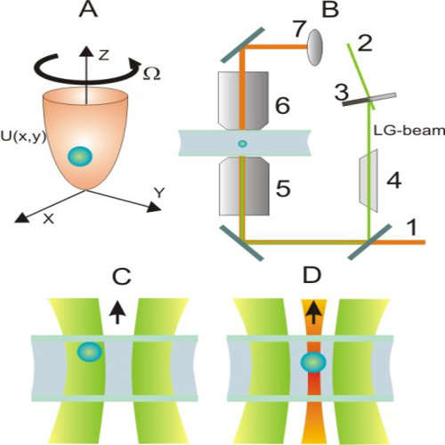
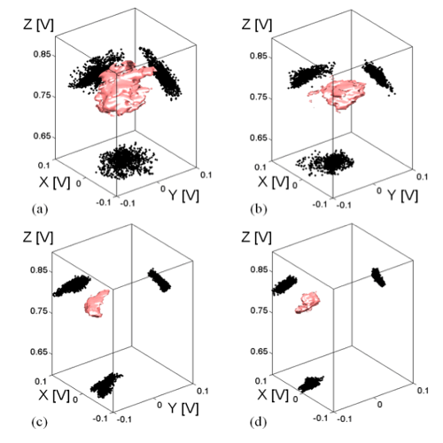
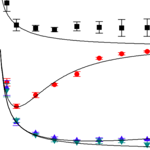
Usually in the presence of a background noise an increased effort put in controlling a system stabilizes its behavior. Rarely it is thought that an increased control of the system can lead to a looser response and, therefore, to a poorer performance. Strikingly there are many systems that show this weird behavior; examples can be drawn form physical, biological, and social systems. Until now no simple and general mechanism underlying such behaviors has been identified. Here we show that such a mechanism, named stochastic resonant damping, can be provided by the interplay between the background noise and the control exerted on the system. We experimentally verify our prediction on a physical model system based on a colloidal particle held in an oscillating optical potential. Our result adds a tool for the study of intrinsically noisy phenomena, joining the many constructive facets of noise identified in the past decades—for example, stochastic resonance, noise-induced activation, and Brownian ratchets.
Surface Plasmon Optical Tweezers published in Phys. Rev. Lett.

Surface plasmon optical tweezers: Tunable optical manipulation in the femtonewton range

Surface plasmon optical tweezers: Tunable optical manipulation in the femtonewton range
Maurizio Righini, Giovanni Volpe, Christian Girard, Dmitri Petrov & Romain Quidant
Physical Review Letters 100(18), 186804 (2008)
DOI: 10.1103/PhysRevLett.100.186804

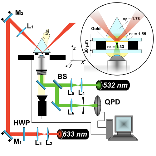
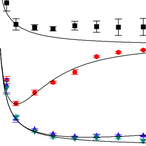
We present a quantitative analysis of 2D surface plasmon based optical tweezers able to trap microcolloids at a patterned metal surface under low laser intensity. Photonic force microscopy is used to assess the properties of surface plasmon traps, such as confinement and stiffness, revealing stable trapping with forces in the range of a few tens of femtonewtons. We also investigate the specificities of surface plasmon tweezers with respect to conventional 3D tweezers responsible for their selectivity to the trapped specimen’s size. The accurate engineering of the trapping properties through the adjustment of the illumination parameters opens new perspectives in the realization of future optically driven on-a-chip devices.

Singular Point Characterization in Microscopic Flows published in Phys. Rev. E

Singular point characterization in microscopic flows

Singular point characterization in microscopic flows
Giorgio Volpe, Giovanni Volpe & Dmitri Petrov
Physical Review E 77(3), 037301 (2008)
DOI: 10.1103/PhysRevE.77.037301
arXiv: 0711.0923

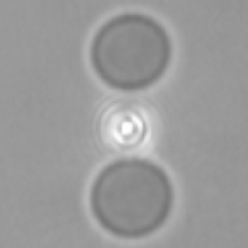
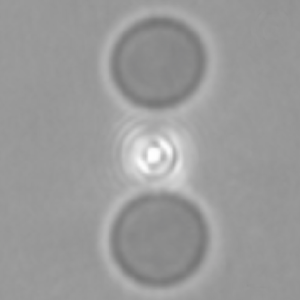
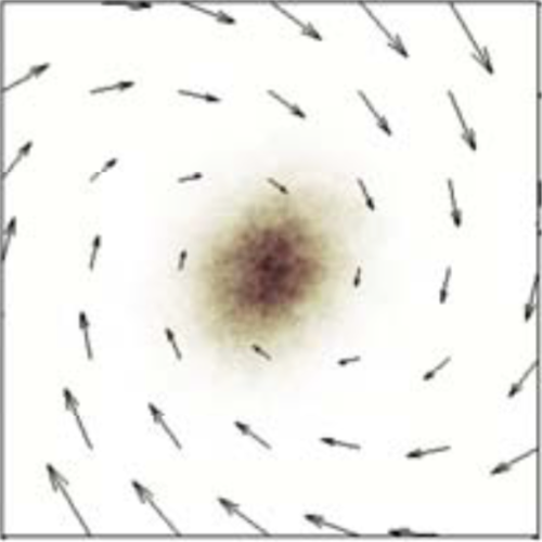
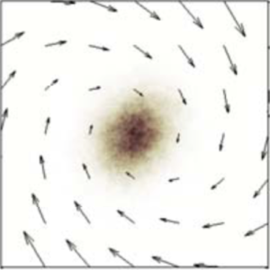
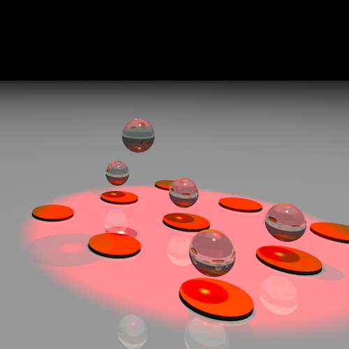
We suggest an approach to microrheology based on optical traps capable of measuring fluid fluxes around singular points of fluid flows. We experimentally demonstrate this technique, applying it to the characterization of controlled flows produced by a set of birefringent spheres spinning due to the transfer of light angular momentum. Unlike the previous techniques, this method is able to distinguish between a singular point in a complex flow and the absence of flow at all; furthermore it permits us to characterize the stability of the singular point.

Photonics Torque Microscopy published in Phys. Rev. E

Brownian motion in a non-homogeneous force field and photonic force microscope

Brownian motion in a non-homogeneous force field and photonic force microscope
Giorgio Volpe, Giovanni Volpe & Dmitri Petrov
Physical Review E 76(6), 061118 (2007)
DOI: 10.1103/PhysRevE.76.061118
arXiv: 0711.0923

The photonic force microscope (PFM) is an opto-mechanical technique that uses an optically trapped probe to measure forces in the range of pico to femto Newton. For a correct use of the PFM, the force field has to be homogeneous on the scale of the Brownian motion of the trapped probe. This condition implicates that the force field must be conservative, excluding the possibility of a rotational component. However, there are cases where these assumptions are not fulfilled. Here, we show how to expand the PFM technique in order to deal with these cases. We introduce the theory of this enhanced PFM and we propose a concrete analysis workflow to reconstruct the force field from the experimental time series of the probe position. Furthermore, we experimentally verify some particularly important cases, namely, the case of a conservative and of a rotational force field.

Back-scattering Position Detection for PFM published in J. Appl. Phys.

Back-scattering position detection for photonic force microscopy

Back-scattering position detection for photonic force microscopy
Giovanni Volpe, Gregory Kozyreff & Dmitri Petrov
Journal of Applied Physics 102(8), 084701 (2007)
DOI: 10.1063/1.2799047

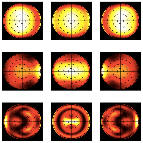
An optically trapped particle is an extremely sensitive probe for the measurement of pico- and femto-Newton forces between the particle and its environment in microscopic systems (photonic force microscopy). A typical setup comprises an optical trap, which holds the probe, and a position sensing system, which uses the scattering of a beam illuminating the probe. Usually the position is accurately determined by measuring the deflection of the forward-scattered light transmitted through the probe. However, geometrical constraints may prevent access to this side of the trap, forcing one to make use of the backscattered light instead. A theory is presented together with numerical results that describes the use of the backscattered light for position detection. With a Mie–Debye approach, we compute the total (incident plus scattered) field and follow its evolution as it is collected by the condenser lenses and projected onto the position detectors and the responses of position sensitive detectors and quadrant photodetectors to the displacement of the probe in the optical trap, both in forward and backward configurations. We find out that in the case of backward detection, for both types of detectors the displacement sensitivity can change sign as a function of the probe size and is null for some critical sizes. In addition, we study the influence of the numerical aperture of the detection system, polarization, and the cross talk between position measurements in orthogonal directions. We finally discuss how these features should be taken into account in experimental designs.

Actin-cytoskeleton Depolymerisation Detection in a Single Cell published in Opt. Express

Real-time actin-cytoskeleton depolymerization detection in a single cell using optical tweezers

Real-time actin-cytoskeleton depolymerization detection in a single cell using optical tweezers
Anna Chiara de Luca, Giovanni Volpe, Anna Morales Drets, Maria Isabel Geli, Giuseppe Pesce, Giulia Rusciano, Antonio Sasso & Dmitri Petrov
Optics Express 15(13), 7922—7932 (2007)
DOI: 10.1364/OE.15.007922

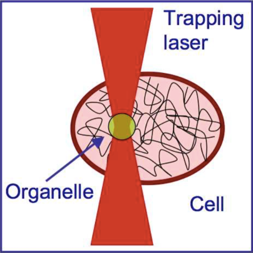
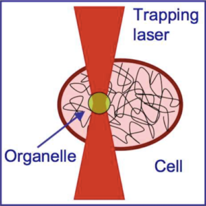
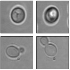
The cytoskeleton provides the backbone structure for the cellular organization, determining, in particular, the cellular mechanical properties. These are important factors in many biological processes, as, for instance, the metastatic process of malignant cells. In this paper, we demonstrate the possibility of monitoring the cytoskeleton structural transformations in optically trapped yeast cells (Saccharomyces cerevisiae) by tracking the forward scattered light via a quadrant photodiode. We distinguished normal cells from cells treated with latrunculin A, a drug which is known to induce the actin-cytoskeleton depolymerization. Since the proposed technique relies only on the inherent properties of the optical trap, without requiring external markers or biochemical sensitive spectroscopic techniques, it can be readily combined with existing optical tweezers setups.

Torque Detection using Brownian Fluctuations published in Phys. Rev. Lett.

Torque detection using Brownian fluctuations

Torque detection using Brownian fluctuations
Giovanni Volpe & Dmitri Petrov
Physical Review Letters 97(21), 210603 (2006)
DOI: 10.1103/PhysRevLett.97.210603

We report the statistical analysis of the movement of a submicron particle confined in a harmonic potential in the presence of a torque. The absolute value of the torque can be found from the auto- and cross-correlation functions of the particle’s coordinates. We experimentally prove this analysis by detecting the torque produced onto an optically trapped particle by an optical beam with orbital angular momentum.

Also featured in “Focus: Hidden Twists and Turns”, Physics 18, 17 (December 1, 2006)

Surface Plasmon Radiation Forces published in Phys. Rev. Lett.

Surface plasmon radiation forces

Surface plasmon radiation forces
Giovanni Volpe, Romain Quidant, Gonçal Badenes & Dmitri Petrov
Physical Review Letters 96(23), 238101 (2006)
DOI: 10.1103/PhysRevLett.96.238101

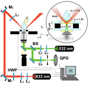
We report the first experimental observation of momentum transfer from a surface plasmon to a single dielectric sphere. Using a photonic force microscope, we measure the plasmon radiation forces on different polystyrene beads as a function of their distance from the metal surface. We show that the force magnitude at resonance is strongly enhanced compared to a nonresonant illumination. Measurements performed as a function of the probe particle size indicate that optical manipulation by plasmon fields has a strong potential for optical sorting.

Dynamics of a Growing Cell in an Optical Trap published in Appl. Phys. Lett.

Dynamics of a growing cell in an optical trap

Dynamics of a growing cell in an optical trap
Giovanni Volpe, Gajendra Pratap Singh & Dmitri Petrov
Applied Physics Letters 88(23), 231106 (2006)
DOI: 10.1063/1.2213015

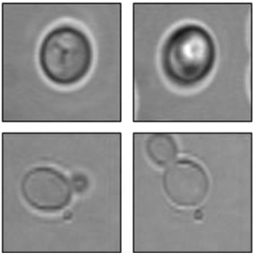
We analyze the forward scattered light from a single optically trapped cellduring its growth. We show that the cell continues adjusting itself to the applied optical force because of the growth processes, and hence it keeps changing its orientation in the trap. We point out the importance of taking this variation into account in the interpretation of spectroscopic data. This method can also be used as a means for cell identification and cell sorting.

Monitoring Yeast Cell Growth Using Raman published in J. Raman Spectrosc.

The lag phase and G1 phase of a single yeast cell monitored by Raman microspectroscopy

The lag phase and G1 phase of a single yeast cell monitored by Raman microspectroscopy
Gajendra P. Singh, Giovanni Volpe, Caitriona M. Creely, Helga Grötsch, Isabel M. Geli & Dmitri Petrov
Journal of Raman Spectroscopy 37(8), 858—864 (2006)
DOI: 10.1002/jrs.1520

We optically trapped a single yeast cell for up to 3 h and monitored the changes in the Raman spectra during the lag phase of its growth and the G1 phase of its cell cycle. A non‐budding cell (corresponding either to the G0 or G1 phase) was chosen for each experiment. During the lag phase, the cell synthesises new proteins and lipids and the observed behaviour of the peaks corresponding to these constituents as well as those of RNA served as a sensitive indicator of the adaptation of the cell to its changed environment. Temporal behaviour of the Raman peaks observed was different in the lag phase as compared to the late lag phase. Two different laser wavelengths were applied to study the effect of long‐term optical trapping on the living cells. Yeast cells killed either by boiling or by a chemical protocol were also trapped for a long time in a single beam optical trap to understand the effect of optical trapping on the behaviour of observed Raman peaks. The changes observed in the Raman spectra of a trapped yeast cell in the late G1 phase or the beginning of S phase corresponded to the growth of a bud.