Amyloid Network Topology in Alzheimer published in Cerebral Cortex

Amyloid network topology characterizes the progression of Alzheimer’s disease during the predementia stages

Amyloid network topology characterizes the progression of Alzheimer’s disease during the predementia stages
Joana B. Pereira, Tor Olof Strandberg, Sebastian Palmqvist, Giovanni Volpe, Danielle van Westen, Eric Westman & Oskar Hansson, for the Alzheimer’s Disease Neuroimaging Initiative
Cerebral Cortex 28(1), 340—349 (2018)
DOI: 10.1093/cercor/bhx294

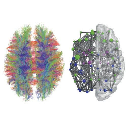
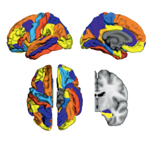
There is increasing evidence showing that the accumulation of the amyloid-β (Aβ) peptide into extracellular plaques is a central event in Alzheimer’s disease (AD). These abnormalities can be detected as lowered levels of Aβ42 in the cerebrospinal fluid (CSF) and are followed by increased amyloid burden on positron emission tomography (PET) several years before the onset of dementia. The aim of this study was to assess amyloid network topology in nondemented individuals with early stage Aβ accumulation, defined as abnormal CSF Aβ42 levels and normal Florbetapir PET (CSF+/PET−), and more advanced Aβ accumulation, defined as both abnormal CSF Aβ42 and Florbetapir PET (CSF+/PET+). The amyloid networks were built using correlations in the mean 18F-florbetapir PET values between 72 brain regions and analyzed using graph theory analyses. Our findings showed an association between early amyloid stages and increased covariance as well as shorter paths between several brain areas that overlapped with the default-mode network (DMN). Moreover, we found that individuals with more advanced amyloid accumulation showed more widespread changes in brain regions both within and outside the DMN. These findings suggest that amyloid network topology could potentially be used to assess disease progression in the predementia stages of AD.

Abnormal Structural Brain Connectome in Preclinical Alzheimer published in Cerebral Cortex

Abnormal structural brain connectome in individuals with preclinical Alzheimer’s disease

Abnormal structural brain connectome in individuals with preclinical Alzheimer’s disease
Joana B. Pereira, Danielle van Westen, Erik Stomrud, Tor Olof Strandberg, Giovanni Volpe, Eric Westman & Oskar Hansson
Cerebral Cortex 28(10), 3638—3649 (2018)
DOI: 10.1093/cercor/bhx236

Alzheimer’s disease has a long preclinical phase during which amyloid pathology and neurodegeneration accumulate in the brain without producing overt cognitive deficits. It is currently unclear whether these early disease stages are associated with a progressive disruption in the communication between brain regions that subsequently leads to cognitive decline and dementia. In this study we assessed the organization of structural networks in cognitively normal (CN) individuals harboring amyloid pathology (A+N−), neurodegeneration (A−N+), or both (A+N+) from the prospective and longitudinal Swedish BioFINDER study. We combined graph theory with diffusion tensor imaging to investigate integration, segregation, and centrality measures in the brain connectome in the previous groups. At baseline, our findings revealed a disrupted network topology characterized by longer paths, lower efficiency, increased clustering and modularity in CN A−N+ and CN A+N+, but not in CN A+N−. After 2 years, CN A+N+ showed significant abnormalities in all global network measures, whereas CN A−N+ only showed abnormalities in the global efficiency. Network connectivity and organization were associated with memory in CN A+N+ individuals. Altogether, our findings suggest that amyloid pathology is not sufficient to disrupt structural network topology, whereas neurodegeneration is.


Featured in “Nuke med helps diagnose early Alzheimer’s from amyloid network topology”, HealthImaging, 14 Nov 2017