Sex differences in multilayer functional network topology over the course of aging in 37543 UK Biobank participants accepted on Network Neuroscience

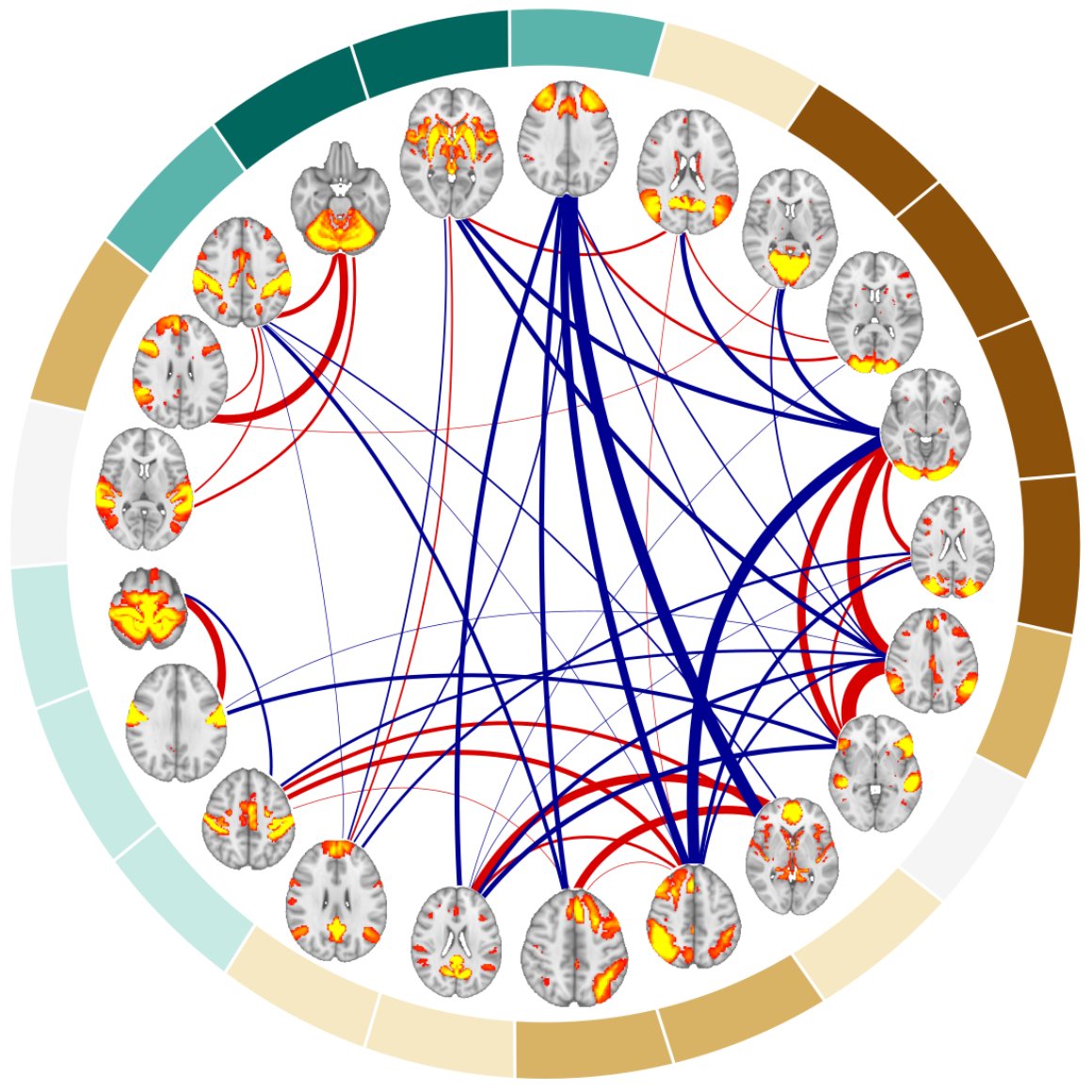
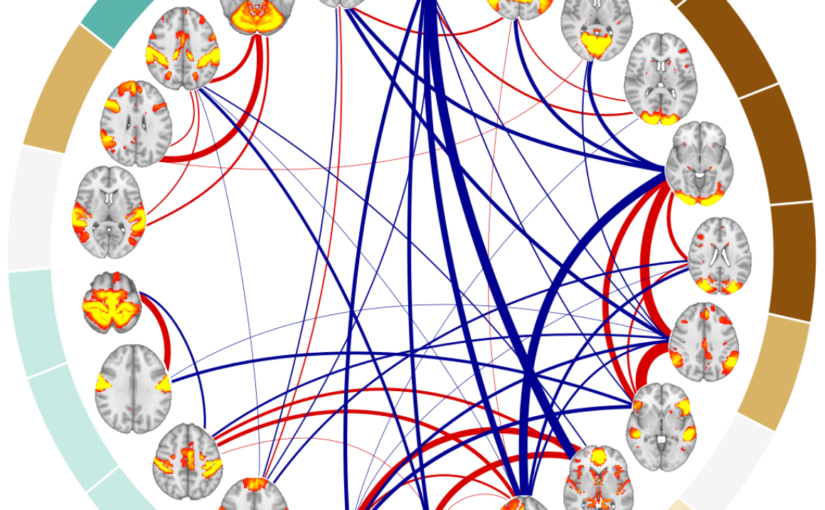
Example of the 21 resting-state networks used as nodes and their positive (red) and negative connections (blue) for one of 140 the subjects included in the analyses. (Image by the Authors of the manuscript.)
Sex differences in multilayer functional network topology over the course of aging in 37543 UK Biobank participants
Mite Mijalkov, Dániel Veréb, Oveis Jamialahmadi, Anna Canal-Garcia, Emiliano Gómez-Ruiz, Didac Vidal-Piñeiro, Stefano Romeo, Giovanni Volpe, Joana B. Pereira
Network Neuroscience 1-40 (2022)
doi: 10.1162/netn_a_00286
medRxiv: 10.1101/2022.03.08.22272089

Aging is a major risk factor for cardiovascular and neurodegenerative disorders, with considerable societal and economic implications. Healthy aging is accompanied by changes in functional connectivity between and within resting-state functional networks, which have been associated with cognitive decline. However, there is no consensus on the impact of sex on these age-related functional trajectories. Here, we show that multilayer measures provide crucial information on the interaction between sex and age on network topology, allowing for better assessment of cognitive, structural, and cardiovascular risk factors that have been shown to differ between men and women, as well as providing additional insights into the genetic influences on changes in functional connectivity that occur during aging. In a large cross-sectional sample of 37543 individuals from the UK Biobank cohort, we demonstrate that such multilayer measures that capture the relationship between positive and negative connections are more sensitive to sex-related changes in the whole-brain connectivity patterns and their topological architecture throughout aging, when compared to standard connectivity and topological measures. Our findings indicate that multilayer measures contain previously unknown information on the relationship between sex and age, which opens up new avenues for research into functional brain connectivity in aging.
Neural Network Training with Highly Incomplete Datasets published in Machine Learning: Science and Technology

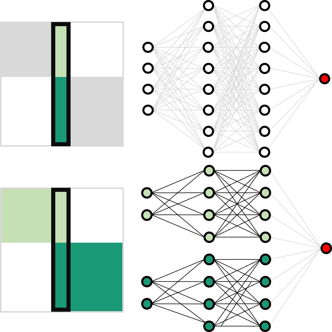
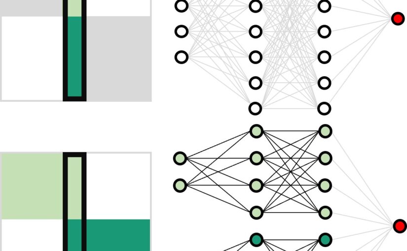
Working principles for training neural networks with highly incomplete dataset: vanilla (upper panel) vs GapNet (lower panel) (Image by Yu-Wei Chang.)
Neural Network Training with Highly Incomplete Datasets
Yu-Wei Chang, Laura Natali, Oveis Jamialahmadi, Stefano Romeo, Joana B. Pereira, Giovanni Volpe
Machine Learning: Science and Technology 3, 035001 (2022)
arXiV: 2107.00429
doi: 10.1088/2632-2153/ac7b69

Neural network training and validation rely on the availability of large high-quality datasets. However, in many cases only incomplete datasets are available, particularly in health care applications, where each patient typically undergoes different clinical procedures or can drop out of a study. Since the data to train the neural networks need to be complete, most studies discard the incomplete datapoints, which reduces the size of the training data, or impute the missing features, which can lead to artefacts. Alas, both approaches are inadequate when a large portion of the data is missing. Here, we introduce GapNet, an alternative deep-learning training approach that can use highly incomplete datasets. First, the dataset is split into subsets of samples containing all values for a certain cluster of features. Then, these subsets are used to train individual neural networks. Finally, this ensemble of neural networks is combined into a single neural network whose training is fine-tuned using all complete datapoints. Using two highly incomplete real-world medical datasets, we show that GapNet improves the identification of patients with underlying Alzheimer’s disease pathology and of patients at risk of hospitalization due to Covid-19. By distilling the information available in incomplete datasets without having to reduce their size or to impute missing values, GapNet will permit to extract valuable information from a wide range of datasets, benefiting diverse fields from medicine to engineering.

Press release on Extracting quantitative biological information from bright-field cell images using deep learning

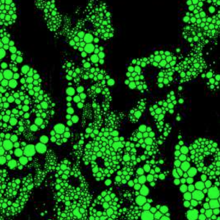
Virtually-stained generated image for lipid-droplet.

The article Extracting quantitative biological information from bright-field cell images using deep learning has been featured in a press release of the University of Gothenburg.

The study, recently published in Biophysics Reviews, shows how artificial intelligence can be used to develop faster, cheaper and more reliable information about cells, while also eliminating the disadvantages from using chemicals in the process.

Here the links to the press releases on Cision:
Swedish: Effektivare studier av celler med ny AI-metod
English: More effective cell studies using new AI method

Here the links to the press releases in the News of the University of Gothenburg:
Swedish: Effektivare studier av celler med ny AI-metod
English: More effective cell studies using new AI method

Extracting quantitative biological information from brightfield cell images using deep learning featured in AIP SciLight

The article Extracting quantitative biological information from brightfield cell images using deep learning
has been featured in: “Staining Cells Virtually Offers Alterative Approach to Chemical Dyes”, AIP SciLight (July 23, 2021).

Scilight showcases the most interesting research across the physical sciences published in AIP Publishing Journals.

Scilight is published weekly (52 issues per year) by AIP Publishing.

Extracting quantitative biological information from bright-field cell images using deep learning published in Biophysics Reviews

Virtually-stained generated image for lipid-droplet.
Extracting quantitative biological information from bright-field cell images using deep learning
Saga Helgadottir, Benjamin Midtvedt, Jesús Pineda, Alan Sabirsh, Caroline B. Adiels, Stefano Romeo, Daniel Midtvedt, Giovanni Volpe
Biophysics Rev. 2, 031401 (2021)
arXiv: 2012.12986
doi: 10.1063/5.0044782

Quantitative analysis of cell structures is essential for biomedical and pharmaceutical research. The standard imaging approach relies on fluorescence microscopy, where cell structures of interest are labeled by chemical staining techniques. However, these techniques are often invasive and sometimes even toxic to the cells, in addition to being time-consuming, labor-intensive, and expensive. Here, we introduce an alternative deep-learning-powered approach based on the analysis of bright-field images by a conditional generative adversarial neural network (cGAN). We show that this approach can extract information from the bright-field images to generate virtually-stained images, which can be used in subsequent downstream quantitative analyses of cell structures. Specifically, we train a cGAN to virtually stain lipid droplets, cytoplasm, and nuclei using bright-field images of human stem-cell-derived fat cells (adipocytes), which are of particular interest for nanomedicine and vaccine development. Subsequently, we use these virtually-stained images to extract quantitative measures about these cell structures. Generating virtually-stained fluorescence images is less invasive, less expensive, and more reproducible than standard chemical staining; furthermore, it frees up the fluorescence microscopy channels for other analytical probes, thus increasing the amount of information that can be extracted from each cell.

Diagnosis of a genetic disease improves with machine learning, a summary in Swedish published in Fysikaktuellt

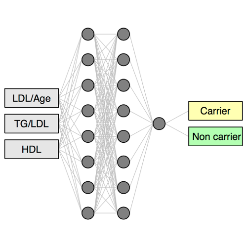
Neural networks consist of a series of connected layers of neurons, whose connection weights are adjusted to learn how to determine the diagnosis from the input data.

A summary in Swedish of our previously published article “Virtual genetic diagnosis for familial hypercholesterolemia powered by machine learning” has been published in Fysikaktuellt, the journal of the Swedish Physical Society (Svenska fysikersamfundet).

Article: “Diagnostisering av sjukdomar förbättras med maskininlärning”, Saga Helgadottir, Giovanni Volpe and Stefano Romeo (in Swedish)

Original article: Virtual genetic diagnosis for familial hypercholesterolemia powered by machine learning

Press release: 
Algoritm lär sig diagnostisera genetisk sjukdom (in Swedish)
An algorithm that learns to diagnose genetic disease (in English)

Virtual genetic diagnosis for familial hypercholesterolemia powered by machine learning published in European Journal of Preventive Cardiology

Neural networks consist of a series of connected layers of neurons, whose connection weights are adjusted to learn how to determine the diagnosis from the input data.

Virtual genetic diagnosis for familial hypercholesterolemia powered by machine learning
Anna Pina, Saga Helgadottir, Rosellina Margherita Mancina, Chiara Pavanello, Carlo Pirazzi, Tiziana Montalcini, Roberto Henriques, Laura Calabresi, Olov Wiklund, M Paula Macedo, Luca Valenti, Giovanni Volpe, Stefano Romeo
European Journal of Preventive Cardiology (2020)
doi: https://doi.org/10.1177/2047487319898951

Aims

Familial hypercholesterolemia (FH) is the most common genetic disorder of lipid metabolism. The gold standard for FH diagnosis is genetic testing, available, however, only in selected university hospitals. Clinical scores – for example, the Dutch Lipid Score – are often employed as alternative, more accessible, albeit less accurate FH diagnostic tools. The aim of this study is to obtain a more reliable approach to FH diagnosis by a “virtual” genetic test using machine-learning approaches.

Methods and results

We used three machine-learning algorithms (a classification tree (CT), a gradient boosting machine (GBM), a neural network (NN)) to predict the presence of FH-causative genetic mutations in two independent FH cohorts: the FH Gothenburg cohort (split into training data (N = 174) and internal test (N = 74)) and the FH-CEGP Milan cohort (external test, N = 364). By evaluating their area under the receiver operating characteristic (AUROC) curves, we found that the three machine-learning algorithms performed better (AUROC 0.79 (CT), 0.83 (GBM), and 0.83 (NN) on the Gothenburg cohort, and 0.70 (CT), 0.78 (GBM), and 0.76 (NN) on the Milan cohort) than the clinical Dutch Lipid Score (AUROC 0.68 and 0.64 on the Gothenburg and Milan cohorts, respectively) in predicting carriers of FH-causative mutations.

Conclusion

In the diagnosis of FH-causative genetic mutations, all three machine-learning approaches we have tested outperform the Dutch Lipid Score, which is the clinical standard. We expect these machine-learning algorithms to provide the tools to implement a virtual genetic test of FH. These tools might prove particularly important for lipid clinics without access to genetic testing.