Medical image segmentation using deep learning.
Hang Zhao
3 May 2022, 11:00
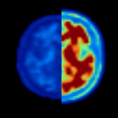
Image segmentation and synthesis of CT image based on deep learning: Deep learning methods for medical image segmentation are hindered by the lack of training data. This thesis aims to develop a method that overcomes this problem. Basic U-net trained on XCAT phantom data was tested first. The segmentation results were unsatisfactory even when artificial quantum noise was added. As a workaround, CycleGAN was used to add tissue textures to the XCAT phantom images by analyzing patient CT images. The generated images were used to train the network. The textures introduced by CycleGAN improved the segmentation, but some errors remained. Basic U-net was replaced with Attention U-net, which further improved the segmentation. More work is needed to fine-tune and thoroughly evaluate the method. The results obtained so far demonstrate the potential of this method for the segmentation of medical images. The proposed algorithms may be used in iterative image reconstruction algorithms in multi-energy computed tomography.
3D Cell nuclei segmentation using digital nuclei phantom and 3D deep learning methods : The analysis of microscopy image is helpful to pathological analysis. Nowadays, deep learning has shown the capabilities of processing the medical imaging data. However, developing deep learning methods in microscopy image analysis can be challenging because of the lack of ground truth and various resolution of microscopy image data. This project aims to build a digital nuclei phantom that simulates the actual microscopy images, including mitotic rate, nucleus size, noise, point spread function, and diverse resolutions. The phantom images were used to train 3D deep neural network for nuclei segmentation. The trained neural network was tested for segmentation on datasets with different resolutions. The neural network successfully performed segmentation on most resolutions in our dataset, and the segmentation results reflect the morphology and density of nuclei in microscopy images. The future work will focus on improving the nuclei phantom to generate more realistic phantom images, thereby further helping with segmentation.
Bio:
My name is Hang, I took my bachelor’s in China on radiophysics, and master’s at Linköping University on biomedical imaging. After I graduate, I joined Karolinska as a research assistant on 3D microscopy image processing. Now, I am working at Linköping university as a full time research engineer, contributing to cardiovascular MR image processing, supervised by Petter Dyverfeldt.