Monitoring Yeast Cell Growth Using Raman published in J. Raman Spectrosc.

The lag phase and G1 phase of a single yeast cell monitored by Raman microspectroscopy

The lag phase and G1 phase of a single yeast cell monitored by Raman microspectroscopy
Gajendra P. Singh, Giovanni Volpe, Caitriona M. Creely, Helga Grötsch, Isabel M. Geli & Dmitri Petrov
Journal of Raman Spectroscopy 37(8), 858—864 (2006)
DOI: 10.1002/jrs.1520

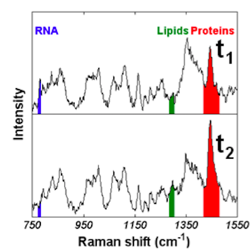
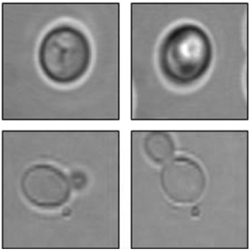
We optically trapped a single yeast cell for up to 3 h and monitored the changes in the Raman spectra during the lag phase of its growth and the G1 phase of its cell cycle. A non‐budding cell (corresponding either to the G0 or G1 phase) was chosen for each experiment. During the lag phase, the cell synthesises new proteins and lipids and the observed behaviour of the peaks corresponding to these constituents as well as those of RNA served as a sensitive indicator of the adaptation of the cell to its changed environment. Temporal behaviour of the Raman peaks observed was different in the lag phase as compared to the late lag phase. Two different laser wavelengths were applied to study the effect of long‐term optical trapping on the living cells. Yeast cells killed either by boiling or by a chemical protocol were also trapped for a long time in a single beam optical trap to understand the effect of optical trapping on the behaviour of observed Raman peaks. The changes observed in the Raman spectra of a trapped yeast cell in the late G1 phase or the beginning of S phase corresponded to the growth of a bud.

Raman Imaging of Floating Cells published in Opt. Express

Raman imaging of floating cells

Raman imaging of floating cells
Caitriona M. Creely, Giovanni Volpe, Gajendra P. Singh, Marta Soler & Dmitri Petrov
Optics Express 13(16), 6105–6110 (2005)
DOI: 10.1364/OPEX.13.006105

Raman imaging can yield spatially resolved biochemical information from living cells. To date there have been no Raman images published of cells in suspension because of the problem of immobilising them suitably to acquire space-resolved spectra. In this paper in order to overcome this problem the use of holographic optical tweezers is proposed and implemented, and data is shown for spatially resolved Raman spectroscopy of a live cell in suspension.

Detection of Hyperosmotic Stress by Raman published in Anal. Chem.

Real-time detection of hyperosmotic stress response in optically trapped single yeast cells using Raman microspectroscopy.

Real-time detection of hyperosmotic stress response in optically trapped single yeast cells using Raman microspectroscopy
Gajendra P. Singh, Caitriona M. Creely, Giovanni Volpe, Helga Grötsch & Dmitri Petrov
Analytical Chemistry 77(8), 2564–2568 (2005)
DOI: 10.1021/ac048359j

Living cells survive environmentally stressful conditions by initiating a stress response. We monitored changes in the Raman spectra of optically trapped Saccharomyces cerevisiae yeast cell under normal, heat-treated, and hyperosmotic stress conditions. It is shown that when glucose was used to exert hyperosmotic stress, two chemical substancesglycerol and ethanolcan be monitored in real time in a single cell.