Deep learning in light–matter interactions published in Nanophotonics

Artificial neurons can be combined in a dense neural network (DNN), where the input layer is connected to the output layer via a set of hidden layers. (Image by the Authors.)
Deep learning in light–matter interactions
Daniel Midtvedt, Vasilii Mylnikov, Alexander Stilgoe, Mikael Käll, Halina Rubinsztein-Dunlop and Giovanni Volpe
Nanophotonics, 11(14), 3189-3214 (2022)
doi: 10.1515/nanoph-2022-0197

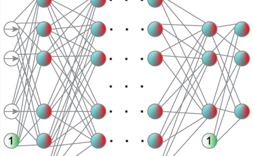
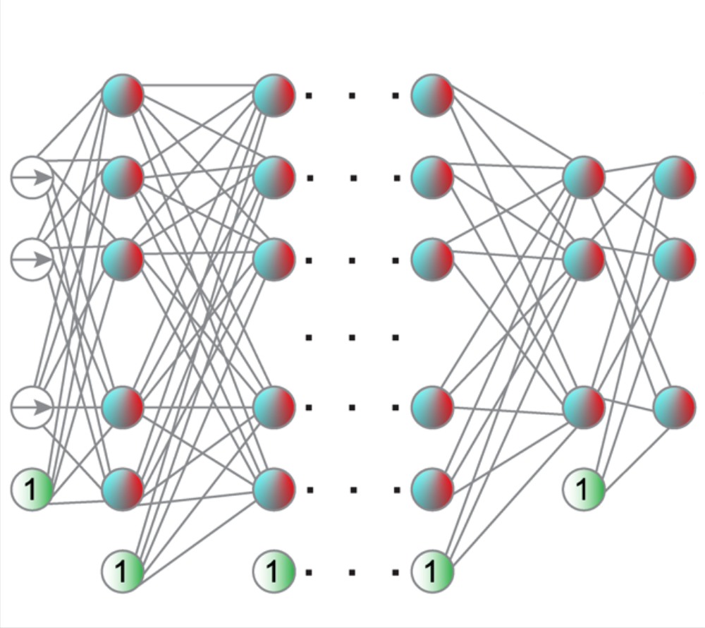
The deep-learning revolution is providing enticing new opportunities to manipulate and harness light at all scales. By building models of light–matter interactions from large experimental or simulated datasets, deep learning has already improved the design of nanophotonic devices and the acquisition and analysis of experimental data, even in situations where the underlying theory is not sufficiently established or too complex to be of practical use. Beyond these early success stories, deep learning also poses several challenges. Most importantly, deep learning works as a black box, making it difficult to understand and interpret its results and reliability, especially when training on incomplete datasets or dealing with data generated by adversarial approaches. Here, after an overview of how deep learning is currently employed in photonics, we discuss the emerging opportunities and challenges, shining light on how deep learning advances photonics.

Label-free nanofluidic scattering microscopy of size and mass of single diffusing molecules and nanoparticles published in Nature Methods

Kymographs of DNA inside Channel II. (Image by the Authors.)
Label-free nanofluidic scattering microscopy of size and mass of single diffusing molecules and nanoparticles
Barbora Špačková, Henrik Klein Moberg, Joachim Fritzsche, Johan Tenghamn, Gustaf Sjösten, Hana Šípová-Jungová, David Albinsson, Quentin Lubart, Daniel van Leeuwen, Fredrik Westerlund, Daniel Midtvedt, Elin K. Esbjörner, Mikael Käll, Giovanni Volpe & Christoph Langhammer
Nature Methods 19, 751–758 (2022)
doi: 10.1038/s41592-022-01491-6

Label-free characterization of single biomolecules aims to complement fluorescence microscopy in situations where labeling compromises data interpretation, is technically challenging or even impossible. However, existing methods require the investigated species to bind to a surface to be visible, thereby leaving a large fraction of analytes undetected. Here, we present nanofluidic scattering microscopy (NSM), which overcomes these limitations by enabling label-free, real-time imaging of single biomolecules diffusing inside a nanofluidic channel. NSM facilitates accurate determination of molecular weight from the measured optical contrast and of the hydrodynamic radius from the measured diffusivity, from which information about the conformational state can be inferred. Furthermore, we demonstrate its applicability to the analysis of a complex biofluid, using conditioned cell culture medium containing extracellular vesicles as an example. We foresee the application of NSM to monitor conformational changes, aggregation and interactions of single biomolecules, and to analyze single-cell secretomes.

Directed Brain Connectivity Identifies Widespread Functional Network Abnormalities in Parkinson’s Disease published in Cerebral Cortex

Visual display of the nodes that show significant differences between controls and participants with PD in network measures using the anti-symmetric correlation method. (Image by the Authors.)
Directed Brain Connectivity Identifies Widespread Functional Network Abnormalities in Parkinson’s Disease
Mite Mijalkov, Giovanni Volpe, Joana B Pereira
Cerebral Cortex 32(3), 593–607 (2022)
doi: 10.1093/cercor/bhab237

Parkinson’s disease (PD) is a neurodegenerative disorder characterized by topological abnormalities in large-scale functional brain networks, which are commonly analyzed using undirected correlations in the activation signals between brain regions. This approach assumes simultaneous activation of brain regions, despite previous evidence showing that brain activation entails causality, with signals being typically generated in one region and then propagated to other ones. To address this limitation, here, we developed a new method to assess whole-brain directed functional connectivity in participants with PD and healthy controls using antisymmetric delayed correlations, which capture better this underlying causality. Our results show that whole-brain directed connectivity, computed on functional magnetic resonance imaging data, identifies widespread differences in the functional networks of PD participants compared with controls, in contrast to undirected methods. These differences are characterized by increased global efficiency, clustering, and transitivity combined with lower modularity. Moreover, directed connectivity patterns in the precuneus, thalamus, and cerebellum were associated with motor, executive, and memory deficits in PD participants. Altogether, these findings suggest that directional brain connectivity is more sensitive to functional network differences occurring in PD compared with standard methods, opening new opportunities for brain connectivity analysis and development of new markers to track PD progression.

Multiplex Connectome Changes across the Alzheimer’s Disease Spectrum Using Gray Matter and Amyloid Data published in Cerebral Cortex

Brain nodes. (Image taken from the article.)
Multiplex Connectome Changes across the Alzheimer’s Disease Spectrum Using Gray Matter and Amyloid Data
Mite Mijalkov, Giovanni Volpe, Joana B Pereira
Anna Canal-Garcia, Emiliano Gómez-Ruiz, Mite Mijalkov, Yu-Wei Chang, Giovanni Volpe, Joana B Pereira, Alzheimer’s Disease Neuroimaging Initiative
Cerebral Cortex, bhab429 (2022)
doi: 10.1093/cercor/bhab429

The organization of the Alzheimer’s disease (AD) connectome has been studied using graph theory using single neuroimaging modalities such as positron emission tomography (PET) or structural magnetic resonance imaging (MRI). Although these modalities measure distinct pathological processes that occur in different stages in AD, there is evidence that they are not independent from each other. Therefore, to capture their interaction, in this study we integrated amyloid PET and gray matter MRI data into a multiplex connectome and assessed the changes across different AD stages. We included 135 cognitively normal (CN) individuals without amyloid-β pathology (Aβ−) in addition to 67 CN, 179 patients with mild cognitive impairment (MCI) and 132 patients with AD dementia who all had Aβ pathology (Aβ+) from the Alzheimer’s Disease Neuroimaging Initiative. We found widespread changes in the overlapping connectivity strength and the overlapping connections across Aβ-positive groups. Moreover, there was a reorganization of the multiplex communities in MCI Aβ + patients and changes in multiplex brain hubs in both MCI Aβ + and AD Aβ + groups. These findings offer a new insight into the interplay between amyloid-β pathology and brain atrophy over the course of AD that moves beyond traditional graph theory analyses based on single brain networks.

Comparison of Two-Dimensional- and Three-Dimensional-Based U-Net Architectures for Brain Tissue Classification in One-Dimensional Brain CT published in Frontiers of Computational Neuroscience

CT is split into smaller patches. (Image by the Authors.)
Comparison of Two-Dimensional- and Three-Dimensional-Based U-Net Architectures for Brain Tissue Classification in One-Dimensional Brain CT
Meera Srikrishna, Rolf A. Heckemann, Joana B. Pereira, Giovanni Volpe, Anna Zettergren, Silke Kern, Eric Westman, Ingmar Skoog and Michael Schöll
Frontiers of Computational Neuroscience 15, 785244 (2022)
doi: 10.3389/fncom.2021.785244

Brain tissue segmentation plays a crucial role in feature extraction, volumetric quantification, and morphometric analysis of brain scans. For the assessment of brain structure and integrity, CT is a non-invasive, cheaper, faster, and more widely available modality than MRI. However, the clinical application of CT is mostly limited to the visual assessment of brain integrity and exclusion of copathologies. We have previously developed two-dimensional (2D) deep learning-based segmentation networks that successfully classified brain tissue in head CT. Recently, deep learning-based MRI segmentation models successfully use patch-based three-dimensional (3D) segmentation networks. In this study, we aimed to develop patch-based 3D segmentation networks for CT brain tissue classification. Furthermore, we aimed to compare the performance of 2D- and 3D-based segmentation networks to perform brain tissue classification in anisotropic CT scans. For this purpose, we developed 2D and 3D U-Net-based deep learning models that were trained and validated on MR-derived segmentations from scans of 744 participants of the Gothenburg H70 Cohort with both CT and T1-weighted MRI scans acquired timely close to each other. Segmentation performance of both 2D and 3D models was evaluated on 234 unseen datasets using measures of distance, spatial similarity, and tissue volume. Single-task slice-wise processed 2D U-Nets performed better than multitask patch-based 3D U-Nets in CT brain tissue classification. These findings provide support to the use of 2D U-Nets to segment brain tissue in one-dimensional (1D) CT. This could increase the application of CT to detect brain abnormalities in clinical settings.

Raman Tweezers for Tire and Road Wear Micro- and Nanoparticles Analysis published in Environmental Science: Nano

Optical beam focused into the liquid: the tire particles are pushed away from the laser focus.

Raman Tweezers for Tire and Road Wear Micro- and Nanoparticles Analysis
Pietro Giuseppe Gucciardi, Gillibert Raymond, Alessandro Magazzù, Agnese Callegari, David Bronte Ciriza, Foti Antonino, Maria Grazia Donato, Onofrio M. Maragò, Giovanni Volpe, Marc Lamy de La Chapelle & Fabienne Lagarde
Environmental Science: Nano 9, 145 – 161 (2022)
ChemRxiv: https://doi.org/10.33774/chemrxiv-2021-h59n1
doi: https://doi.org/10.1039/D1EN00553G

Tire and Road Wear Particles (TRWP) are non-exhaust particulate matter generated by road transport means during the mechanical abrasion of tires, brakes and roads. TRWP accumulate on the roadsides and are transported into the aquatic ecosystem during stormwater runoffs. Due to their size (sub-millimetric) and rubber content (elastomers), TRWP are considered microplastics (MPs). While the amount of the MPs polluting the water ecosystem with sizes from ~ 5 μm to more than 100 μm is known, the fraction of smaller particles is unknown due to the technological gap in the detection and analysis of < 5 μm MPs. Here we show that Raman Tweezers, a combination of optical tweezers and Raman spectroscopy, can be used to trap and chemically analyze individual TWRPs in a liquid environment, down to the sub-micrometric scale. Using tire particles mechanically grinded from aged car tires in water solutions, we show that it is possible to optically trap individual sub-micron particles, in a so-called 2D trapping configuration, and acquire their Raman spectrum in few tens of seconds. The analysis is then extended to samples collected from a brake test platform, where we highlight the presence of sub-micrometric agglomerates of rubber and brake debris, thanks to the presence of additional spectral features other than carbon. Our results show the potential of Raman Tweezers in environmental pollution analysis and highlight the formation of nanosized TRWP during wear.

Featured in:
University of Gothenburg > News and Events: New technology enables the detection of microplastics from road wear
Phys.org > News > Nanotechnology:New technology enables the detection of microplastics from road wear
Nonsologreen > Green: Le Raman-tweezers per la guerra alle nanoplastiche che inquinano fiumi e mari

Objective comparison of methods to decode anomalous diffusion published in Nature Communications

An illustration of anomalous diffusion. (Image by Gorka Muñoz-Gil.)
Objective comparison of methods to decode anomalous diffusion
Gorka Muñoz-Gil, Giovanni Volpe, Miguel Angel Garcia-March, Erez Aghion, Aykut Argun, Chang Beom Hong, Tom Bland, Stefano Bo, J. Alberto Conejero, Nicolás Firbas, Òscar Garibo i Orts, Alessia Gentili, Zihan Huang, Jae-Hyung Jeon, Hélène Kabbech, Yeongjin Kim, Patrycja Kowalek, Diego Krapf, Hanna Loch-Olszewska, Michael A. Lomholt, Jean-Baptiste Masson, Philipp G. Meyer, Seongyu Park, Borja Requena, Ihor Smal, Taegeun Song, Janusz Szwabiński, Samudrajit Thapa, Hippolyte Verdier, Giorgio Volpe, Arthur Widera, Maciej Lewenstein, Ralf Metzler, and Carlo Manzo
Nat. Commun. 12, Article number: 6253 (2021)
doi: 10.1038/s41467-021-26320-w
arXiv: 2105.06766

Deviations from Brownian motion leading to anomalous diffusion are found in transport dynamics from quantum physics to life sciences. The characterization of anomalous diffusion from the measurement of an individual trajectory is a challenging task, which traditionally relies on calculating the trajectory mean squared displacement. However, this approach breaks down for cases of practical interest, e.g., short or noisy trajectories, heterogeneous behaviour, or non-ergodic processes. Recently, several new approaches have been proposed, mostly building on the ongoing machine-learning revolution. To perform an objective comparison of methods, we gathered the community and organized an open competition, the Anomalous Diffusion challenge (AnDi). Participating teams applied their algorithms to a commonly-defined dataset including diverse conditions. Although no single method performed best across all scenarios, machine-learning-based approaches achieved superior performance for all tasks. The discussion of the challenge results provides practical advice for users and a benchmark for developers.

Active droploids published in Nature Communications

Active droploids. (Image taken from the article.)
Active droploids
Jens Grauer, Falko Schmidt, Jesús Pineda, Benjamin Midtvedt, Hartmut Löwen, Giovanni Volpe & Benno Liebchen
Nat. Commun. 12, 6005 (2021)
doi: 10.1038/s41467-021-26319-3
arXiv: 2109.10677

Active matter comprises self-driven units, such as bacteria and synthetic microswimmers, that can spontaneously form complex patterns and assemble into functional microdevices. These processes are possible thanks to the out-of-equilibrium nature of active-matter systems, fueled by a one-way free-energy flow from the environment into the system. Here, we take the next step in the evolution of active matter by realizing a two-way coupling between active particles and their environment, where active particles act back on the environment giving rise to the formation of superstructures. In experiments and simulations we observe that, under light-illumination, colloidal particles and their near-critical environment create mutually-coupled co-evolving structures. These structures unify in the form of active superstructures featuring a droplet shape and a colloidal engine inducing self-propulsion. We call them active droploids—a portmanteau of droplet and colloids. Our results provide a pathway to create active superstructures through environmental feedback.

The environment topography alters the transition from single-cell populations to multicellular structures in Myxococcus xanthus published in Science Advances

M. xanthus cell-cell and cell-particle local interactions during cellular aggregation.
The environment topography alters the transition from single-cell populations to multicellular structures in Myxococcus xanthus
Karla C. Hernández Ramos, Edna Rodríguez-Sánchez, Juan Antonio Arias del Angel, Alejandro V. Arzola, Mariana Benítez, Ana E. Escalante, Alessio Franci, Giovanni Volpe, Natsuko Rivera-Yoshida
Sci. Adv. 7(35), eabh2278 (2021)
bioRxiv: 10.1101/2021.01.27.428527
doi: 10.1126/sciadv.abh2278

The social soil-dwelling bacteria Myxococcus xanthus can form multicellular structures, known as fruiting bodies. Experiments in homogeneous environments have shown that this process is affected by the physico-chemical properties of the substrate, but they have largely neglected the role of complex topographies. We experimentally demonstrate that the topography alters single-cell motility and multicellular organization in M. xanthus. In topographies realized by randomly placing silica particles over agar plates, we observe that the cells’ interaction with particles drastically modifies the dynamics of cellular aggregation, leading to changes in the number, size and shape of the fruiting bodies, and even to arresting their formation in certain conditions. We further explore this type of cell-particle interaction in a minimal computational model. These results provide fundamental insights into how the environment topography influences the emergence of complex multicellular structures from single cells, which is a fundamental problem of biological, ecological and medical relevance.

The Cognitive Connectome in Healthy Aging published in Frontiers in Aging Neuroscience

Age-independent cognitive connectome in the whole cohort.
The Cognitive Connectome in Healthy Aging
Eloy Garcia-Cabello, Lissett Gonzalez-Burgos, Joana B. Pereira, Juan Andres Hernández-Cabrera, Eric Westman, Giovanni Volpe, José Barroso, & Daniel Ferreira
Front. Aging Neurosci. 13, 530 (2021)
doi: 10.3389/fnagi.2021.694254

Objectives: Cognitive aging has been extensively investigated using both univariate and multivariate analyses. Sophisticated multivariate approaches such as graph theory could potentially capture unknown complex associations between multiple cognitive variables. The aim of this study was to assess whether cognition is organized into a structure that could be called the “cognitive connectome,” and whether such connectome differs between age groups.

Methods: A total of 334 cognitively unimpaired individuals were stratified into early-middle-age (37–50 years, n = 110), late-middle-age (51–64 years, n = 106), and elderly (65–78 years, n = 118) groups. We built cognitive networks from 47 cognitive variables for each age group using graph theory and compared the groups using different global and nodal graph measures.

Results: We identified a cognitive connectome characterized by five modules: verbal memory, visual memory—visuospatial abilities, procedural memory, executive—premotor functions, and processing speed. The elderly group showed reduced transitivity and average strength as well as increased global efficiency compared with the early-middle-age group. The late-middle-age group showed reduced global and local efficiency and modularity compared with the early-middle-age group. Nodal analyses showed the important role of executive functions and processing speed in explaining the differences between age groups.

Conclusions: We identified a cognitive connectome that is rather stable during aging in cognitively healthy individuals, with the observed differences highlighting the important role of executive functions and processing speed. We translated the connectome concept from the neuroimaging field to cognitive data, demonstrating its potential to advance our understanding of the complexity of cognitive aging.