 Fast volumetric light-sheet microscopy
Seminar by Omar E. Olarte
from Universidad ECCI, Colombia (and ICFO, Barcelona, Spain)
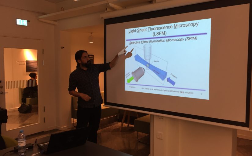
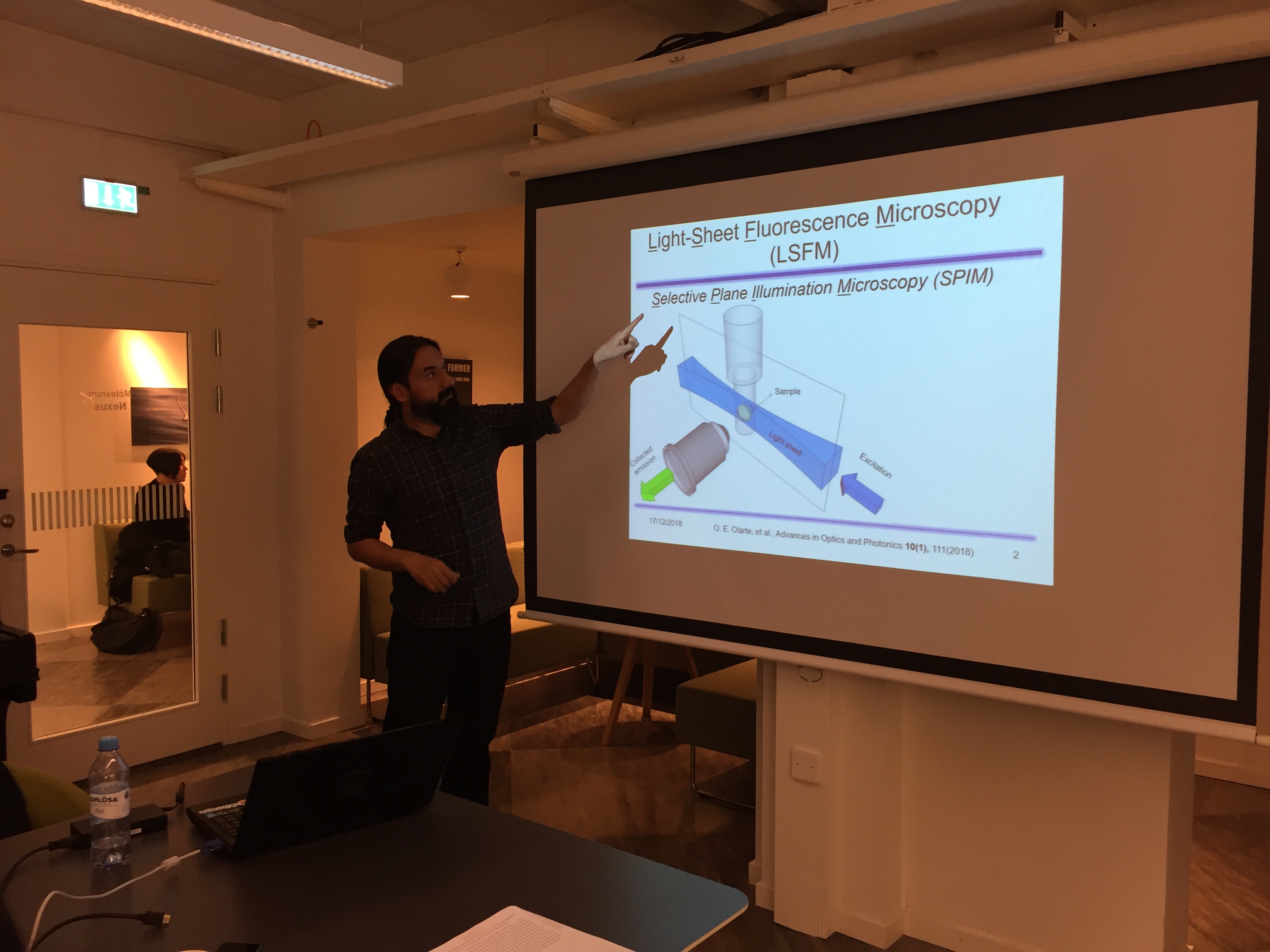
Light sheet fluorescence microscopy (LSFM) is a convenient tool for bio-imaging as it efficiently collects the generated fluorescence while at the same time minimizes photobleaching. For these reasons LSFM, being based on an intrinsic 2D illumination strategy, has been put forward as an interesting candidate for fast volumetric imaging.
We report on a LSFM microscope, combined with the use of wavefront coding (WFC) techniques, for fast volumetric imaging. This provides intrinsic 3D imaging capabilities as it extends the depth of field (DOF) of the microscope. In addition, because of the extended DOF, the light sheet can be axially scanned at fast speeds. As only the light sheet is moved, fast 3D imaging can be achieved without the need of any sample or objective movement. Since typical light scanning devices can run at KHz rates, 3D volumetric acquisition speeds will only be limited by the reading speed of the camera or the required signal to noise ratio.
WFC works at the expenses of introducing a controlled aberration to the system which blurs the resulting images. Then it requires a final deconvolution step to recover the image sharpness that would impose a limitation on the application. Here we present computational tools to perform real-time deconvolution and visualization of the images obtained with WFC-LSFM. To speed up such deconvolution processes, a routine directly developed in a GPUs has been developed. We present preliminary results on realistic computer generated images showing that real-time deconvolution and visualization is possible.
Place: Nexus
Time: 17 December 2018, 16:00