Juan S. Sierra, Jesus Pineda, Daniela Rueda, Alejandro Tello, Angelica M. Prada, Virgilio Galvis, Giovanni Volpe, Maria S. Millan, Lenny A. Romero, Andres G. Marrugo
Biomedical Optics Express 14, 335-351 (2023)
doi: 10.1364/BOE.477495
arXiv: 2210.07102
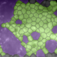
Specular microscopy assessment of the human corneal endothelium (CE) in Fuchs’ dystrophy is challenging due to the presence of dark image regions called guttae. This paper proposes a UNet-based segmentation approach that requires minimal post-processing and achieves reliable CE morphometric assessment and guttae identification across all degrees of Fuchs’ dystrophy. We cast the segmentation problem as a regression task of the cell and gutta signed distance maps instead of a pixel-level classification task as typically done with UNets. Compared to the conventional UNet classification approach, the distance-map regression approach converges faster in clinically relevant parameters. It also produces morphometric parameters that agree with the manually-segmented ground-truth data, namely the average cell density difference of -41.9 cells/mm2 (95% confidence interval (CI) [-306.2, 222.5]) and the average difference of mean cell area of 14.8 um2 (95% CI [-41.9, 71.5]). These results suggest a promising alternative for CE assessment.